Background
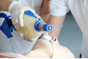
Rapid Sequence Intubation (RSI) is a medical procedure used to secure the airway in patients who are at risk of aspiration or cannot maintain adequate oxygenation or ventilation. It is a highly controlled and time-sensitive technique that minimizes the risk of aspiration by combining rapid administration of a sedative (induction agent) and a neuromuscular blocking agent, allowing for prompt and safe endotracheal intubation.
Indications
Airway Protection
Inability to protect the airway: Due to altered mental status, coma, or neurologic conditions (e.g., stroke, traumatic brain injury).
Risk of aspiration: Present in patients with a full stomach (recent meal, gastrointestinal bleed, or bowel obstruction) or those vomiting.
Oxygenation or Ventilation Failure
Severe hypoxemia: Despite high-flow oxygen or non-invasive ventilation (e.g., ARDS, pneumonia).
Hypercapnia with respiratory acidosis: Typically seen in advanced COPD or other causes of respiratory failure.
Imminent respiratory arrest: Fatigue or failure of respiratory muscles in conditions like status asthmaticus or myasthenia gravis.
Anticipated Clinical Deterioration
Trauma patients with airway compromise: Facial trauma, neck hematoma, or burns causing swelling.
Anaphylaxis or angioedema: Involving airway structures, leading to potential obstruction.
Smoke inhalation injuries: That may cause airway edema.
Neurologic Indications
Increased intracranial pressure (ICP): To prevent secondary brain injury and facilitate hyperventilation if indicated.
Seizures: Refractory status epilepticus requiring airway protection and sedation.
Contraindications
Known Allergy to RSI Medications:
Allergy or hypersensitivity to any of the medications planned for induction or paralysis (e.g., succinylcholine, etomidate, or rocuronium).
Severe Anticipated Difficulty with Airway Management:
Patients with a high likelihood of failed intubation and where alternative airway strategies (e.g., awake intubation or surgical airway) may be safer.
Severe Cardiovascular Instability:
Hemodynamic instability or conditions where induction agents could worsen hypotension, making the risks outweigh the benefits without prior optimization.
Difficult Airway Anticipation:
Trauma, swelling, tumors, or anatomical abnormalities. Ensure airway adjuncts (e.g., video laryngoscopy, bougie, cricothyrotomy kits) and skilled personnel are available.
Increased Risk of Aspiration:
Severe gastrointestinal bleeding or active vomiting where preoxygenation and suctioning may need modification.
Outcomes
Equipment
Laryngoscope
Endotracheal Tubes (ETT)
Stylet
Supraglottic Airway Devices (SGA)
Bag-Valve-Mask (BVM)
Oxygen Source
Nasal Cannula
Patient Preparation
Medication Preparation
Premedication (optional, based on clinical situation):
Atropine: For bradycardia prevention in pediatrics.
Lidocaine: For suspected intracranial hypertension or reactive airway disease.
Fentanyl: For blunting hemodynamic response.
Induction Agent (choose based on patient condition):
Etomidate: Hemodynamically stable patients.
Ketamine: Hypotension or bronchospasm.
Propofol: Contraindicated in hypotension but preferred in stable patients.
Neuromuscular Blocker:
Succinylcholine: Rapid onset, short duration (contraindicated in hyperkalemia, crush injuries, or burns).
Rocuronium: Alternative with longer duration.
Patient position:
Place the patient in the sniffing position for adequate visualization; flex the neck and extend the head. This position helps to align the axes and facilitates visualization of the glottic opening.
Technique
Step 1-Preparation:
Equipment:
Airway tools: Laryngoscope (with appropriate blade), endotracheal tubes (ETT) of different sizes, stylet, bougie.
Oxygenation: Bag-valve mask (BVM), high-flow nasal cannula, and suction.
Monitoring: Pulse oximetry, ECG, capnography, and blood pressure monitoring.
Medications: Induction agent (e.g., etomidate, propofol, ketamine), paralytic agent (e.g., succinylcholine, rocuronium), and emergency drugs (e.g., epinephrine, atropine).
IV access: Ensure functional and patent IV/IO lines.
Step 2-Pre-intubation assessment:
Evaluate airway using tools like LEMON (Look externally, Evaluate 3-3-2, Mallampati score, Obstruction/Obesity, Neck mobility).
Predict difficulty with bag-mask ventilation, laryngoscopy, and intubation.
Step 3-Preoxygenation:
To increase oxygen reserves and delay hypoxia during apnea.
Provide 100% oxygen using a non-rebreather mask, BVM, or high-flow nasal cannula for 3–5 minutes if possible.
In emergencies, perform 4–8 large tidal breaths with 100% oxygen.
Step 4-Pre-treatment (Optional):
Consider administering pre-treatment drugs for specific indications:
Lidocaine: To blunt intracranial pressure (ICP) rise in head injury.
Fentanyl: To mitigate sympathetic response in cases like hypertension.
Atropine: In pediatric patients to prevent bradycardia from vagal stimulation.
Step 5-Induction:
Administer the induction agent (e.g., etomidate for hemodynamic stability, ketamine for bronchodilation in asthma, or propofol for rapid onset but with caution in hypotension).
Onset: Agents typically work within 30–60 seconds.
Step 6-Paralysis with Sedation:
Administer a paralytic immediately after the induction agent:
Succinylcholine: Rapid onset (30–60 seconds) but contraindicated in hyperkalemia, burns, crush injuries, and neuromuscular diseases.
Rocuronium: Non-depolarizing alternative with a slightly slower onset (45–90 seconds).
Step 7-Positioning:
Align the airway axes (oral, pharyngeal, and laryngeal) using the “sniffing position”:
Extend the neck and elevate the head.
For obese patients, use a ramped position with the external auditory meatus aligned to the sternal notch.
Ensure the suction device is readily available and functional.
Step 8-Intubation:

Rapid sequence intubation
Perform direct or video laryngoscopy:
Visualize the glottis and vocal cords.
Pass the endotracheal tube (ETT) through the cords until the cuff is just beyond the cords.
Inflate the ETT cuff with air to achieve a seal.
Confirm placement using end-tidal CO₂ (capnography), chest rise, and auscultation (bilateral breath sounds).
Step 9-Confirmation:
Ensure proper tube placement:
End-tidal CO₂ detection (gold standard): A persistent waveform indicates proper placement.
Symmetric chest rise and breath sounds.
No gastric insufflation sounds (auscultate over the epigastrium).
Secure the ETT and note the depth at the lips (usually 20–24 cm in adults).
Step 10-Post-intubation Care:
Provide mechanical ventilation or BVM support.
Monitor vital signs and oxygenation closely.
Adjust ventilator settings as needed.
Consider a post-intubation chest X-ray to confirm tube depth and rule out complications like pneumothorax or mainstem intubation.
Complications
Hypoxia:
Insufficient pre-oxygenation or prolonged intubation attempts can lead to oxygen deprivation.
Patients with pre-existing respiratory or cardiovascular compromise are at higher risk.
Aspiration:
Even with cricoid pressure, the risk of aspiration of gastric contents remains, especially in patients with a full stomach.
Esophageal Intubation:
Incorrect placement of the endotracheal tube can lead to ineffective ventilation and hypoxia.
Must be identified promptly using capnography and auscultation.
Trauma:
Physical injury to the lips, teeth, tongue, pharynx, vocal cords, or trachea during intubation.
More likely in patients with difficult anatomy or poor visualization.
Hemodynamic Instability:
Induction agents (e.g., etomidate, propofol) can cause hypotension.
Sympatholytic effects of medications or vagal stimulation may exacerbate instability.
Laryngospasm or Bronchospasm:
May occur during induction or intubation, especially in patients with reactive airway diseases.

Rapid Sequence Intubation (RSI) is a medical procedure used to secure the airway in patients who are at risk of aspiration or cannot maintain adequate oxygenation or ventilation. It is a highly controlled and time-sensitive technique that minimizes the risk of aspiration by combining rapid administration of a sedative (induction agent) and a neuromuscular blocking agent, allowing for prompt and safe endotracheal intubation.
Airway Protection
Inability to protect the airway: Due to altered mental status, coma, or neurologic conditions (e.g., stroke, traumatic brain injury).
Risk of aspiration: Present in patients with a full stomach (recent meal, gastrointestinal bleed, or bowel obstruction) or those vomiting.
Oxygenation or Ventilation Failure
Severe hypoxemia: Despite high-flow oxygen or non-invasive ventilation (e.g., ARDS, pneumonia).
Hypercapnia with respiratory acidosis: Typically seen in advanced COPD or other causes of respiratory failure.
Imminent respiratory arrest: Fatigue or failure of respiratory muscles in conditions like status asthmaticus or myasthenia gravis.
Anticipated Clinical Deterioration
Trauma patients with airway compromise: Facial trauma, neck hematoma, or burns causing swelling.
Anaphylaxis or angioedema: Involving airway structures, leading to potential obstruction.
Smoke inhalation injuries: That may cause airway edema.
Neurologic Indications
Increased intracranial pressure (ICP): To prevent secondary brain injury and facilitate hyperventilation if indicated.
Seizures: Refractory status epilepticus requiring airway protection and sedation.
Known Allergy to RSI Medications:
Allergy or hypersensitivity to any of the medications planned for induction or paralysis (e.g., succinylcholine, etomidate, or rocuronium).
Severe Anticipated Difficulty with Airway Management:
Patients with a high likelihood of failed intubation and where alternative airway strategies (e.g., awake intubation or surgical airway) may be safer.
Severe Cardiovascular Instability:
Hemodynamic instability or conditions where induction agents could worsen hypotension, making the risks outweigh the benefits without prior optimization.
Difficult Airway Anticipation:
Trauma, swelling, tumors, or anatomical abnormalities. Ensure airway adjuncts (e.g., video laryngoscopy, bougie, cricothyrotomy kits) and skilled personnel are available.
Increased Risk of Aspiration:
Severe gastrointestinal bleeding or active vomiting where preoxygenation and suctioning may need modification.
Laryngoscope
Endotracheal Tubes (ETT)
Stylet
Supraglottic Airway Devices (SGA)
Bag-Valve-Mask (BVM)
Oxygen Source
Nasal Cannula
Patient Preparation
Medication Preparation
Premedication (optional, based on clinical situation):
Atropine: For bradycardia prevention in pediatrics.
Lidocaine: For suspected intracranial hypertension or reactive airway disease.
Fentanyl: For blunting hemodynamic response.
Induction Agent (choose based on patient condition):
Etomidate: Hemodynamically stable patients.
Ketamine: Hypotension or bronchospasm.
Propofol: Contraindicated in hypotension but preferred in stable patients.
Neuromuscular Blocker:
Succinylcholine: Rapid onset, short duration (contraindicated in hyperkalemia, crush injuries, or burns).
Rocuronium: Alternative with longer duration.
Patient position:
Place the patient in the sniffing position for adequate visualization; flex the neck and extend the head. This position helps to align the axes and facilitates visualization of the glottic opening.
Step 1-Preparation:
Equipment:
Airway tools: Laryngoscope (with appropriate blade), endotracheal tubes (ETT) of different sizes, stylet, bougie.
Oxygenation: Bag-valve mask (BVM), high-flow nasal cannula, and suction.
Monitoring: Pulse oximetry, ECG, capnography, and blood pressure monitoring.
Medications: Induction agent (e.g., etomidate, propofol, ketamine), paralytic agent (e.g., succinylcholine, rocuronium), and emergency drugs (e.g., epinephrine, atropine).
IV access: Ensure functional and patent IV/IO lines.
Step 2-Pre-intubation assessment:
Evaluate airway using tools like LEMON (Look externally, Evaluate 3-3-2, Mallampati score, Obstruction/Obesity, Neck mobility).
Predict difficulty with bag-mask ventilation, laryngoscopy, and intubation.
Step 3-Preoxygenation:
To increase oxygen reserves and delay hypoxia during apnea.
Provide 100% oxygen using a non-rebreather mask, BVM, or high-flow nasal cannula for 3–5 minutes if possible.
In emergencies, perform 4–8 large tidal breaths with 100% oxygen.
Step 4-Pre-treatment (Optional):
Consider administering pre-treatment drugs for specific indications:
Lidocaine: To blunt intracranial pressure (ICP) rise in head injury.
Fentanyl: To mitigate sympathetic response in cases like hypertension.
Atropine: In pediatric patients to prevent bradycardia from vagal stimulation.
Step 5-Induction:
Administer the induction agent (e.g., etomidate for hemodynamic stability, ketamine for bronchodilation in asthma, or propofol for rapid onset but with caution in hypotension).
Onset: Agents typically work within 30–60 seconds.
Step 6-Paralysis with Sedation:
Administer a paralytic immediately after the induction agent:
Succinylcholine: Rapid onset (30–60 seconds) but contraindicated in hyperkalemia, burns, crush injuries, and neuromuscular diseases.
Rocuronium: Non-depolarizing alternative with a slightly slower onset (45–90 seconds).
Step 7-Positioning:
Align the airway axes (oral, pharyngeal, and laryngeal) using the “sniffing position”:
Extend the neck and elevate the head.
For obese patients, use a ramped position with the external auditory meatus aligned to the sternal notch.
Ensure the suction device is readily available and functional.
Step 8-Intubation:

Rapid sequence intubation
Perform direct or video laryngoscopy:
Visualize the glottis and vocal cords.
Pass the endotracheal tube (ETT) through the cords until the cuff is just beyond the cords.
Inflate the ETT cuff with air to achieve a seal.
Confirm placement using end-tidal CO₂ (capnography), chest rise, and auscultation (bilateral breath sounds).
Step 9-Confirmation:
Ensure proper tube placement:
End-tidal CO₂ detection (gold standard): A persistent waveform indicates proper placement.
Symmetric chest rise and breath sounds.
No gastric insufflation sounds (auscultate over the epigastrium).
Secure the ETT and note the depth at the lips (usually 20–24 cm in adults).
Step 10-Post-intubation Care:
Provide mechanical ventilation or BVM support.
Monitor vital signs and oxygenation closely.
Adjust ventilator settings as needed.
Consider a post-intubation chest X-ray to confirm tube depth and rule out complications like pneumothorax or mainstem intubation.
Complications
Hypoxia:
Insufficient pre-oxygenation or prolonged intubation attempts can lead to oxygen deprivation.
Patients with pre-existing respiratory or cardiovascular compromise are at higher risk.
Aspiration:
Even with cricoid pressure, the risk of aspiration of gastric contents remains, especially in patients with a full stomach.
Esophageal Intubation:
Incorrect placement of the endotracheal tube can lead to ineffective ventilation and hypoxia.
Must be identified promptly using capnography and auscultation.
Trauma:
Physical injury to the lips, teeth, tongue, pharynx, vocal cords, or trachea during intubation.
More likely in patients with difficult anatomy or poor visualization.
Hemodynamic Instability:
Induction agents (e.g., etomidate, propofol) can cause hypotension.
Sympatholytic effects of medications or vagal stimulation may exacerbate instability.
Laryngospasm or Bronchospasm:
May occur during induction or intubation, especially in patients with reactive airway diseases.

Both our subscription plans include Free CME/CPD AMA PRA Category 1 credits.

On course completion, you will receive a full-sized presentation quality digital certificate.
A dynamic medical simulation platform designed to train healthcare professionals and students to effectively run code situations through an immersive hands-on experience in a live, interactive 3D environment.

When you have your licenses, certificates and CMEs in one place, it's easier to track your career growth. You can easily share these with hospitals as well, using your medtigo app.


