Refractive surgery is a specialized field in ophthalmology, where various vision impairments are treated including myopia, hyperopia, astigmatism, presbyopia. The primary purpose of the surgery is to diminish or reduce the dependence on glasses or contact lenses through the alteration of corneal tissue or through the intervention of devices for focusing the eye. The corrective surgery mainly employs advanced lasers to either reshape the cornea or change it inner surface. Through surgical refracting the cornea or implantation of artificial lens, the light that enters the eye focuses on the retina wall, enhancing vision.
Refractive surgery
Indications
Myopia (Near-sightedness): Patients with myopia, where distant objects appear blurry, may be candidates if their prescription is stable and falls within the surgical range. Hyperopia, or farsighted: If nearby objects are blurry, they may be candidates for this if the hyperopia is mild to moderate. Astigmatism: It is a condition in which the cornea is irregularly shaped, causing distorted or blurry vision. Refractive surgery can correct this. Presbyopia (Age-Related Farsightedness): Some people have refractive surgery to minimize the need for reading glasses or bifocals, although results may vary, especially for patients over 40. Improvement in Quality of Life: It is the most convenient solution for people who are dependent on glasses or contact lenses.
Contraindications
Severe or Unstable Refractive Error:
High myopia (extreme near-sightedness) or hyperopia (farsightedness) that cannot be safely corrected.
Unstable vision: Changes in vision due to conditions like pregnancy or breastfeeding, or rapidly progressing refractive errors.
Corneal Issues:
Thin corneas: Insufficient corneal thickness to safely perform the procedure.
Irregular corneas: Conditions like keratoconus (a progressive thinning and bulging of the cornea) make refractive surgery risky.
Corneal scars or infections: Any scarring from previous eye injuries or surgeries could impact healing.
Dry Eye Disease:
People with moderate to severe dry eye symptoms may experience worsened symptoms post-surgery.
Eye Diseases and Conditions:
Glaucoma: Increased pressure inside the eye can complicate healing.
Cataracts: If a patient is developing cataracts, they may need to address that first.
Retinal disorders: Conditions like macular degeneration, diabetic retinopathy, or retinal detachment might make surgery unsafe.
Age Considerations:
Under 18 years old: Vision may still be changing, so surgery is generally not recommended until the eyes have stabilized.
Over 40 years old: Age-related changes like presbyopia (difficulty focusing on close objects) can affect the outcome of surgery.
Autoimmune or Healing Disorders:
Conditions like rheumatoid arthritis, lupus, or diabetes can slow healing or interfere with the body’s ability to recover from surgery.
Keloid formation: Tendency to form abnormal scarring after surgery may impact recovery.
Outcomes
Periprocedural care
Equipment
Excimer Laser
Femtosecond Laser
Microkeratome
Wavefront Analyzer
Topographer
Pachymeter
Autorefractor
Laser Cataract Surgery Equipment (for post-surgery needs)
SLT (Scanning Laser Tomography)
Fluorescein Staining & Slit Lamp
Surgical Microscope
Patient preparation
Patient preparation before surgery
Comprehensive Eye Exam
Visual acuity test: For checking the current vision and to find out the severity of refractive error.
Corneal thickness measurement: Using pachymetry for confirming the possibility of performing the surgery because the cornea should be thick enough for the surgery.
Pupil size measurement: With the larger pupils, the side effects of vision such as glare or halos are also likely to occur.
Topography: Computerized corneal mapping to see whether the cornea has a normal shape or not.
Dry eye evaluation: Before refractive surgery intraocular test must be done and dry eye syndrome must be excluded because LASIK surgery causes symptoms of dry eyes much worse.
Refraction: A precise measurement of the refractive error.
Retinal exam: This is done to confirm that there are no underlying conditions that might affect the surgery.
Pre-Operative Counselling
Review of potential risks and complications: These may be dry eyes, glare around lights, halos, under-correction or over-correction.
Realistic expectations: It’s important to set expectations about the potential outcome, which may not result in perfect 20/20 vision, but significant improvement.
Lifestyle and Health Issues
Discontinue certain medications: Certain drugs compromise or enhances the risks of surgery or affects healing processes. In some cases, the patient must temporarily stop their use during the surgery.
Eye makeup: No eye makeup should be worn by a patient for at least 48 hours before the start of the procedure to minimize chances of infection.
Contact lens removal: Soft contact lens users should remove them at least one or two weeks prior the examination.
Individuals who wear hard contact lenses also may have to pull over for 3-4 weeks because these lenses change the surface of the cornea.
Blood thinners: Any patient under anticoagulants will require them to be discontinued as advised by the surgeon before the surgery.
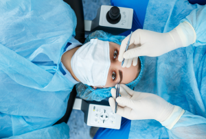
Patient position
Supine Position: Patients are usually placed in a supine position on an operating table.
Head Stabilization: A headrest or a head clamp is employed to fix the head of the patient, at least during the process of implementing this method.
Fixation Light: The patient is requested to stare at a fixation lamp or some different point or target. This aids in eye alignment and helps sustain concentration all through the surgery.
Eye Positioning: The position of the eye is maintained using a speculum to keep the eyelids open, and the patient cannot blink. Sometimes cornea is reshaped or there may be a need to change its position within the eye thus there will be slight shift in position.
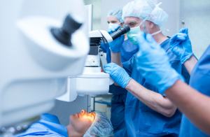
Technique
The most common technique is LASIK (Laser-Assisted in Situ Keratomileusis), although other options like PRK (Photorefractive Keratectomy) and SMILE (Small Incision Lenticule Extraction) are also used.
LASIK Surgery:
Step 1-Preoperative Evaluation: Eye assessment that includes obtaining the central corneal thickness, intraocular pressure, and pupils’ diameter.
Assessing for other factors which might hinder healing of the eyes such as dry eye disorders.
The patient’s vision summary is checked, and, if necessary, the eyes are dilated for better assessment for ophthalmologist.
Step 2-Numbing the Eyes: Medicated Eye drops are instilled so that patient can not feel any pain throughout the procedure.
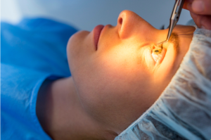
Creating the Corneal Flap: Using either a microkeratome (Fine surgical blade) or more commonly a femtosecond is used to create a thin, hinged flap in the cornea (the clear, front part of the eye).
The local tissue is then gently flapped aside to reveal the corneal layer.
Step 3-Reshaping the Cornea: Application of excimer laser aims at modifying the form of coronal tissue. This laser ablates small amounts of tissue to flatten the cornea for nearsighted patients, or to steepen it for farsighted patients.
The laser used utilizes the parameters such as the specific prescription of the patient and the shape of the cornea.
Step 4-Repositioning the Flap: When the corneal tissue has been reshaped, the flap is put back into position in the exact manner without stitches. Thus, the flap settles neatly in a very natural manner, and the healing of surgery commences.
Step 5-Postoperative Care: Eye drops are given to patients to prevent infection and inflammation of the eye.
The patient is monitored for a short time in the recovery area before being allowed to go home.
Patients usually report better vision within a few hours, and the outcome only improves in the subsequent weeks.
They are followed up to see how the wound is healing and to maximize the potential final visual result.
Complications
Dry Eye Syndrome: The lack of production of tears can occur post-surgery. Most people experience this temporarily, but in some cases, it will persist for longer duration of time. Infection: There is always a chance of infection with any surgical procedure. Though rare, it may cause loss of vision if left untreated. Under correction or Overcorrection: The desired correction may not be achieved. In that case, another surgery or corrective lenses will be needed. Epithelial Ingrowth: In LASIK, cells from the corneal surface can grow into the space under the flap, leading to vision problems. Flap Complications (in LASIK): Issues like incomplete flap creation or dislocation can lead to vision problems or infection. Regression: The eye may gradually return to its pre-surgery refractive state over time, requiring enhancement procedures. Corneal Ectasia: The cornea becomes weakened and bulges outward, a more serious complication, which can lead to severe vision problems. Increased Sensitivity to Light: Some individuals experiences heightened sensitivity to light after surgery.
Refractive surgery is a specialized field in ophthalmology, where various vision impairments are treated including myopia, hyperopia, astigmatism, presbyopia. The primary purpose of the surgery is to diminish or reduce the dependence on glasses or contact lenses through the alteration of corneal tissue or through the intervention of devices for focusing the eye. The corrective surgery mainly employs advanced lasers to either reshape the cornea or change it inner surface. Through surgical refracting the cornea or implantation of artificial lens, the light that enters the eye focuses on the retina wall, enhancing vision.
Refractive surgery
Myopia (Near-sightedness): Patients with myopia, where distant objects appear blurry, may be candidates if their prescription is stable and falls within the surgical range. Hyperopia, or farsighted: If nearby objects are blurry, they may be candidates for this if the hyperopia is mild to moderate. Astigmatism: It is a condition in which the cornea is irregularly shaped, causing distorted or blurry vision. Refractive surgery can correct this. Presbyopia (Age-Related Farsightedness): Some people have refractive surgery to minimize the need for reading glasses or bifocals, although results may vary, especially for patients over 40. Improvement in Quality of Life: It is the most convenient solution for people who are dependent on glasses or contact lenses.
Severe or Unstable Refractive Error:
High myopia (extreme near-sightedness) or hyperopia (farsightedness) that cannot be safely corrected.
Unstable vision: Changes in vision due to conditions like pregnancy or breastfeeding, or rapidly progressing refractive errors.
Corneal Issues:
Thin corneas: Insufficient corneal thickness to safely perform the procedure.
Irregular corneas: Conditions like keratoconus (a progressive thinning and bulging of the cornea) make refractive surgery risky.
Corneal scars or infections: Any scarring from previous eye injuries or surgeries could impact healing.
Dry Eye Disease:
People with moderate to severe dry eye symptoms may experience worsened symptoms post-surgery.
Eye Diseases and Conditions:
Glaucoma: Increased pressure inside the eye can complicate healing.
Cataracts: If a patient is developing cataracts, they may need to address that first.
Retinal disorders: Conditions like macular degeneration, diabetic retinopathy, or retinal detachment might make surgery unsafe.
Age Considerations:
Under 18 years old: Vision may still be changing, so surgery is generally not recommended until the eyes have stabilized.
Over 40 years old: Age-related changes like presbyopia (difficulty focusing on close objects) can affect the outcome of surgery.
Autoimmune or Healing Disorders:
Conditions like rheumatoid arthritis, lupus, or diabetes can slow healing or interfere with the body’s ability to recover from surgery.
Keloid formation: Tendency to form abnormal scarring after surgery may impact recovery.
Equipment
Excimer Laser
Femtosecond Laser
Microkeratome
Wavefront Analyzer
Topographer
Pachymeter
Autorefractor
Laser Cataract Surgery Equipment (for post-surgery needs)
SLT (Scanning Laser Tomography)
Fluorescein Staining & Slit Lamp
Surgical Microscope
Patient preparation
Patient preparation before surgery
Comprehensive Eye Exam
Visual acuity test: For checking the current vision and to find out the severity of refractive error.
Corneal thickness measurement: Using pachymetry for confirming the possibility of performing the surgery because the cornea should be thick enough for the surgery.
Pupil size measurement: With the larger pupils, the side effects of vision such as glare or halos are also likely to occur.
Topography: Computerized corneal mapping to see whether the cornea has a normal shape or not.
Dry eye evaluation: Before refractive surgery intraocular test must be done and dry eye syndrome must be excluded because LASIK surgery causes symptoms of dry eyes much worse.
Refraction: A precise measurement of the refractive error.
Retinal exam: This is done to confirm that there are no underlying conditions that might affect the surgery.
Pre-Operative Counselling
Review of potential risks and complications: These may be dry eyes, glare around lights, halos, under-correction or over-correction.
Realistic expectations: It’s important to set expectations about the potential outcome, which may not result in perfect 20/20 vision, but significant improvement.
Lifestyle and Health Issues
Discontinue certain medications: Certain drugs compromise or enhances the risks of surgery or affects healing processes. In some cases, the patient must temporarily stop their use during the surgery.
Eye makeup: No eye makeup should be worn by a patient for at least 48 hours before the start of the procedure to minimize chances of infection.
Contact lens removal: Soft contact lens users should remove them at least one or two weeks prior the examination.
Individuals who wear hard contact lenses also may have to pull over for 3-4 weeks because these lenses change the surface of the cornea.
Blood thinners: Any patient under anticoagulants will require them to be discontinued as advised by the surgeon before the surgery.
Patient position
Supine Position: Patients are usually placed in a supine position on an operating table.
Head Stabilization: A headrest or a head clamp is employed to fix the head of the patient, at least during the process of implementing this method.
Fixation Light: The patient is requested to stare at a fixation lamp or some different point or target. This aids in eye alignment and helps sustain concentration all through the surgery.
Eye Positioning: The position of the eye is maintained using a speculum to keep the eyelids open, and the patient cannot blink. Sometimes cornea is reshaped or there may be a need to change its position within the eye thus there will be slight shift in position.
The most common technique is LASIK (Laser-Assisted in Situ Keratomileusis), although other options like PRK (Photorefractive Keratectomy) and SMILE (Small Incision Lenticule Extraction) are also used.
LASIK Surgery:
Step 1-Preoperative Evaluation: Eye assessment that includes obtaining the central corneal thickness, intraocular pressure, and pupils’ diameter.
Assessing for other factors which might hinder healing of the eyes such as dry eye disorders.
The patient’s vision summary is checked, and, if necessary, the eyes are dilated for better assessment for ophthalmologist.
Step 2-Numbing the Eyes: Medicated Eye drops are instilled so that patient can not feel any pain throughout the procedure.
Creating the Corneal Flap: Using either a microkeratome (Fine surgical blade) or more commonly a femtosecond is used to create a thin, hinged flap in the cornea (the clear, front part of the eye).
The local tissue is then gently flapped aside to reveal the corneal layer.
Step 3-Reshaping the Cornea: Application of excimer laser aims at modifying the form of coronal tissue. This laser ablates small amounts of tissue to flatten the cornea for nearsighted patients, or to steepen it for farsighted patients.
The laser used utilizes the parameters such as the specific prescription of the patient and the shape of the cornea.
Step 4-Repositioning the Flap: When the corneal tissue has been reshaped, the flap is put back into position in the exact manner without stitches. Thus, the flap settles neatly in a very natural manner, and the healing of surgery commences.
Step 5-Postoperative Care: Eye drops are given to patients to prevent infection and inflammation of the eye.
The patient is monitored for a short time in the recovery area before being allowed to go home.
Patients usually report better vision within a few hours, and the outcome only improves in the subsequent weeks.
They are followed up to see how the wound is healing and to maximize the potential final visual result.
Complications
Dry Eye Syndrome: The lack of production of tears can occur post-surgery. Most people experience this temporarily, but in some cases, it will persist for longer duration of time. Infection: There is always a chance of infection with any surgical procedure. Though rare, it may cause loss of vision if left untreated. Under correction or Overcorrection: The desired correction may not be achieved. In that case, another surgery or corrective lenses will be needed. Epithelial Ingrowth: In LASIK, cells from the corneal surface can grow into the space under the flap, leading to vision problems. Flap Complications (in LASIK): Issues like incomplete flap creation or dislocation can lead to vision problems or infection. Regression: The eye may gradually return to its pre-surgery refractive state over time, requiring enhancement procedures. Corneal Ectasia: The cornea becomes weakened and bulges outward, a more serious complication, which can lead to severe vision problems. Increased Sensitivity to Light: Some individuals experiences heightened sensitivity to light after surgery.
Both our subscription plans include Free CME/CPD AMA PRA Category 1 credits.
Digital Certificate PDF
On course completion, you will receive a full-sized presentation quality digital certificate.
medtigo Simulation
A dynamic medical simulation platform designed to train healthcare professionals and students to effectively run code situations through an immersive hands-on experience in a live, interactive 3D environment.
medtigo Points
medtigo points is our unique point redemption system created to award users for interacting on our site. These points can be redeemed for special discounts on the medtigo marketplace as well as towards the membership cost itself.
Community Forum post/reply = 5 points
*Redemption of points can occur only through the medtigo marketplace, courses, or simulation system. Money will not be credited to your bank account. 10 points = $1.
All Your Certificates in One Place
When you have your licenses, certificates and CMEs in one place, it's easier to track your career growth. You can easily share these with hospitals as well, using your medtigo app.