Background
Retrobulbar block is used for eye surgery where local anesthetic is injected into the space behind the eye.
The injection blocks cranial nerves II, III, VI to prevent eye muscle movement. It blocks ciliary nerves for full ocular sensory anesthesia.
It is indicated to numb the eye, paralyzes their muscles, and block sensation of patient during surgery to prevent any movement.
The retrobulbar space is a cone-shaped area behind the eyeball. The anesthesia is administered through the lower eyelid or outer part of the orbit.
The direction of needle should be towards the apex of the orbit to block the ciliary nerves.
Motor innervations to rectus muscles and sensory fibers from globe are found in cone to nasociliary nerve.
Cranial nerves III and VI pass through the muscle cone, but cranial nerve IV goes outside to innervate the superior oblique muscle.
Globe efferent fibers travel through long and short posterior ciliary nerves to intraconal ciliary ganglion to nasociliary nerve from superior trigeminal division.
Indications
Cataract Surgery
Glaucoma Surgery
Vitrectomy
Retinal Detachment Repair
Strabismus Surgery
Corneal Transplantation
It eliminates the need for general anesthesia to reduce systemic risks.
It provides excellent control of eye movement for accuracy in surgery.
It reduces pain during the procedure with rapid onset of anesthesia.
Contraindications
Outcomes
The retrobulbar block provides an excellent anesthesia for eye and their surrounding area.
Patients experience minimal pain during the procedure due to the effectiveness of numbness that eliminates sensation in eye.
Surgeon ensures that eye remains steady during surgery which is very important in ophthalmic procedures.
The effect of the block disappears within a few hours, but the patient may experience blurred vision.
Equipment required
Patient Preparation
Detailed ocular examination should be done to assess anatomical anomalies.
Check all necessary equipment is available in good condition like local anesthetics, syringes, and needles.
Informed Consent:
Patients should understand procedure, benefits, risks, and alternatives for consent.
Patient Positioning
Patients should be positioned in a supine position with a stable head.
Apply a topical antiseptic around the affected area of the eye.
The patient head should be tilted slightly backward and to opposite side of the injection. 
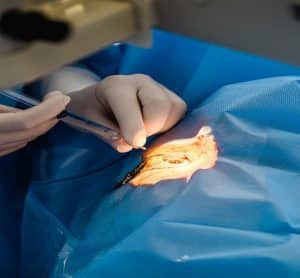
Fig. Retrobulbar block in eye
Technique
Step 1: Landmarks identification:
Mark landmarks between lateral canthus and orbital rim.
Low eyelid palpation of inferior orbital rim allows globe elevation with mild pressure on globe inferior aspect.
Step 2: Insertion of needle:
Insert needle to target retrobulbar space behind eye for medical procedure.
Insert needle to a depth of about 2-3 cm but avoid excessive force to minimize risk of the injury.
Step 3: Administration of injection:
Insert the needle about 1 to 2 mm below lower eyelid margin in conjunctival sac.
Place needle towards the apex of orbit to enter retrobulbar space behind the eye.
Step 4: Removal of needle:
Aspirate to check for blood return. If blood is aspirated withdraw slightly and reorient the needle.
Complications
Retrobulbar Hemorrhage
Ocular Cardiac Reflex
Infection
Globe Perforation
Chemical Toxicity
Eyelid Ecchymosis
Injury to optic nerve due to direct trauma from the needle or hematoma pressure.
A reflex that causes a sudden drop-in heart rate due to pressure on the optic nerve.
Bruising around the eyelid occurs due to bleeding from the injection site.

Retrobulbar block is used for eye surgery where local anesthetic is injected into the space behind the eye.
The injection blocks cranial nerves II, III, VI to prevent eye muscle movement. It blocks ciliary nerves for full ocular sensory anesthesia.
It is indicated to numb the eye, paralyzes their muscles, and block sensation of patient during surgery to prevent any movement.
The retrobulbar space is a cone-shaped area behind the eyeball. The anesthesia is administered through the lower eyelid or outer part of the orbit.
The direction of needle should be towards the apex of the orbit to block the ciliary nerves.
Motor innervations to rectus muscles and sensory fibers from globe are found in cone to nasociliary nerve.
Cranial nerves III and VI pass through the muscle cone, but cranial nerve IV goes outside to innervate the superior oblique muscle.
Globe efferent fibers travel through long and short posterior ciliary nerves to intraconal ciliary ganglion to nasociliary nerve from superior trigeminal division.
Cataract Surgery
Glaucoma Surgery
Vitrectomy
Retinal Detachment Repair
Strabismus Surgery
Corneal Transplantation
It eliminates the need for general anesthesia to reduce systemic risks.
It provides excellent control of eye movement for accuracy in surgery.
It reduces pain during the procedure with rapid onset of anesthesia.
The retrobulbar block provides an excellent anesthesia for eye and their surrounding area.
Patients experience minimal pain during the procedure due to the effectiveness of numbness that eliminates sensation in eye.
Surgeon ensures that eye remains steady during surgery which is very important in ophthalmic procedures.
The effect of the block disappears within a few hours, but the patient may experience blurred vision.
Detailed ocular examination should be done to assess anatomical anomalies.
Check all necessary equipment is available in good condition like local anesthetics, syringes, and needles.
Informed Consent:
Patients should understand procedure, benefits, risks, and alternatives for consent.
Patients should be positioned in a supine position with a stable head.
Apply a topical antiseptic around the affected area of the eye.
The patient head should be tilted slightly backward and to opposite side of the injection. 
Fig. Retrobulbar block in eye
Step 1: Landmarks identification:
Mark landmarks between lateral canthus and orbital rim.
Low eyelid palpation of inferior orbital rim allows globe elevation with mild pressure on globe inferior aspect.
Step 2: Insertion of needle:
Insert needle to target retrobulbar space behind eye for medical procedure.
Insert needle to a depth of about 2-3 cm but avoid excessive force to minimize risk of the injury.
Step 3: Administration of injection:
Insert the needle about 1 to 2 mm below lower eyelid margin in conjunctival sac.
Place needle towards the apex of orbit to enter retrobulbar space behind the eye.
Step 4: Removal of needle:
Aspirate to check for blood return. If blood is aspirated withdraw slightly and reorient the needle.
Retrobulbar Hemorrhage
Ocular Cardiac Reflex
Infection
Globe Perforation
Chemical Toxicity
Eyelid Ecchymosis
Injury to optic nerve due to direct trauma from the needle or hematoma pressure.
A reflex that causes a sudden drop-in heart rate due to pressure on the optic nerve.
Bruising around the eyelid occurs due to bleeding from the injection site.

Both our subscription plans include Free CME/CPD AMA PRA Category 1 credits.

On course completion, you will receive a full-sized presentation quality digital certificate.
A dynamic medical simulation platform designed to train healthcare professionals and students to effectively run code situations through an immersive hands-on experience in a live, interactive 3D environment.

When you have your licenses, certificates and CMEs in one place, it's easier to track your career growth. You can easily share these with hospitals as well, using your medtigo app.


