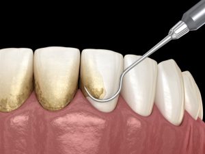
Scaling and Root Planing is a non-surgical procedure to eliminate plaque, tartar, and bacteria from teeth.
Primary treatment for periodontitis targets chronic inflammation in gums, periodontal ligament, and alveolar bone supporting teeth.
Advancements in dental technology made procedures more effective with ultrasonic scalers and better diagnostic tools.
It is designed to eliminate bacterial biofilm and calculus that contribute to gum inflammation. It reduces periodontal pocket depth to prevent further bacterial colonization.
Scaling removes plaque and tartar from the tooth surfaces including below the gumline.
Root Planing smooths out rough areas on the root surfaces to prevent bacteria from reattaching and promote healing.
It acts as therapeutic and preventive measure that reduces inflammation and improves oral health.
Indications
Gingivitis
Periodontitis
Presence of Subgingival Plaque and Calculus
Pre-Surgical Periodontal Therapy
Peri-implant Mucositis
Halitosis
Contraindications
Uncontrolled diabetes mellitus
Uncontrolled hypertension
Severe immunosuppression
Bleeding disorders without proper medical management
Acute Necrotizing Ulcerative Gingivitis
Severe Dental Hypersensitivity
Patients on Anticoagulant or Antiplatelet Therapy
Pregnancy
Severe Anxiety
Outcomes
Decreased inflammation and bleeding from biofilm and calculus removal.
Improved probing pocket depth and gingival tissue reattachment in moderate periodontitis treatments.
Exposed roots can cause sensitivity after plaque removal. Gingival shrinkage post-inflammation may expose roots and affect aesthetics.
Poor oral hygiene risks plaque buildup and recurrent periodontitis without maintenance.
Equipment required
Mouth Mirror
Periodontal Probe
Explorer
Scalers
Curettes
Ultrasonic Scalers
Patient Preparation:
Comprehensive periodontal examination including probing pocket depth, bleeding on probing, and radiographic asses.
Medical history review to identify systemic conditions and medications affecting bleeding.
Educate patients on oral hygiene before procedure.
Informed Consent:
Explain the procedure’s risks and potential complications clearly to the patient.
Patient Positioning:
Patients are positioned in a semi-supine or supine position.
Scaling Procedure
Step 1: Instrumentation Selection
Start with ultrasonic scalers for heavy deposits. Use hand scalers and curettes for precision cleaning.
Step 2: Scaling Movements
Use short and controlled strokes with moderate pressure.
Adapt instrument tip to the tooth surface at approx. 45° to 90° angle.
Work systematically in quadrants to ensure thorough debridement.
Step 3: Frequent Irrigation
Flush pockets with water or antimicrobial agents to reduce bacterial load.
Root Planing Procedure
Step 1: Instrumentation:
Use Gracey curettes with angled at 60° to 70°.
Step 2: Technique:
Apply lighter and longer strokes compared to scaling. Ensure the surface feels smooth and glass-like to prevent bacterial adherence.
Scaling and Root Planing is a non-surgical procedure to eliminate plaque, tartar, and bacteria from teeth.
Primary treatment for periodontitis targets chronic inflammation in gums, periodontal ligament, and alveolar bone supporting teeth.
Advancements in dental technology made procedures more effective with ultrasonic scalers and better diagnostic tools.
It is designed to eliminate bacterial biofilm and calculus that contribute to gum inflammation. It reduces periodontal pocket depth to prevent further bacterial colonization.
Scaling removes plaque and tartar from the tooth surfaces including below the gumline.
Root Planing smooths out rough areas on the root surfaces to prevent bacteria from reattaching and promote healing.
It acts as therapeutic and preventive measure that reduces inflammation and improves oral health.
Gingivitis
Periodontitis
Presence of Subgingival Plaque and Calculus
Pre-Surgical Periodontal Therapy
Peri-implant Mucositis
Halitosis
Uncontrolled diabetes mellitus
Uncontrolled hypertension
Severe immunosuppression
Bleeding disorders without proper medical management
Acute Necrotizing Ulcerative Gingivitis
Severe Dental Hypersensitivity
Patients on Anticoagulant or Antiplatelet Therapy
Pregnancy
Severe Anxiety
Decreased inflammation and bleeding from biofilm and calculus removal.
Improved probing pocket depth and gingival tissue reattachment in moderate periodontitis treatments.
Exposed roots can cause sensitivity after plaque removal. Gingival shrinkage post-inflammation may expose roots and affect aesthetics.
Poor oral hygiene risks plaque buildup and recurrent periodontitis without maintenance.
Mouth Mirror
Periodontal Probe
Explorer
Scalers
Curettes
Ultrasonic Scalers
Patient Preparation:
Comprehensive periodontal examination including probing pocket depth, bleeding on probing, and radiographic asses.
Medical history review to identify systemic conditions and medications affecting bleeding.
Educate patients on oral hygiene before procedure.
Informed Consent:
Explain the procedure’s risks and potential complications clearly to the patient.
Patient Positioning:
Patients are positioned in a semi-supine or supine position.
Step 1: Instrumentation Selection
Start with ultrasonic scalers for heavy deposits. Use hand scalers and curettes for precision cleaning.
Step 2: Scaling Movements
Use short and controlled strokes with moderate pressure.
Adapt instrument tip to the tooth surface at approx. 45° to 90° angle.
Work systematically in quadrants to ensure thorough debridement.
Step 3: Frequent Irrigation
Flush pockets with water or antimicrobial agents to reduce bacterial load.
Root Planing Procedure
Step 1: Instrumentation:
Use Gracey curettes with angled at 60° to 70°.
Step 2: Technique:
Apply lighter and longer strokes compared to scaling. Ensure the surface feels smooth and glass-like to prevent bacterial adherence.
Both our subscription plans include Free CME/CPD AMA PRA Category 1 credits.
Digital Certificate PDF
On course completion, you will receive a full-sized presentation quality digital certificate.
medtigo Simulation
A dynamic medical simulation platform designed to train healthcare professionals and students to effectively run code situations through an immersive hands-on experience in a live, interactive 3D environment.
medtigo Points
medtigo points is our unique point redemption system created to award users for interacting on our site. These points can be redeemed for special discounts on the medtigo marketplace as well as towards the membership cost itself.
Community Forum post/reply = 5 points
*Redemption of points can occur only through the medtigo marketplace, courses, or simulation system. Money will not be credited to your bank account. 10 points = $1.
All Your Certificates in One Place
When you have your licenses, certificates and CMEs in one place, it's easier to track your career growth. You can easily share these with hospitals as well, using your medtigo app.