Background
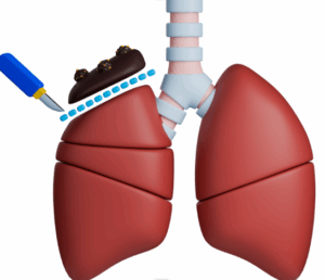
Segmentectomy is a surgical technique used to treat some lung cancers, particularly when the tumor has not grown out of the boundaries of a single section of the lung. It involves removing more bulk lung tissue, including the tumor and the lung segment in which it’s formed.

Segmentectomy
Segmentectomy removes the involved segment of the lung with a margin of healthy tissue surrounding it. Segmentectomy is done at an early stage when the tumor is small, localized, and has not invaded the surrounding tissue, and that’s why it will treat cancer as a curative intervention. Radiological therapy may also be proposed for those who have unsuitable lung function and other health problems, amongst which this type of surgery would not result in effective medical outcomes.
Indications
Early-stage lung cancer: Segmentectomy is an alternative for early-stage Non-small Cell Lung Cancer when it comes to patients with pre-existing lung conditions which make them at high risk for complications if complete lobectomy is carried out.
Lung nodules: If suspected lung nodules are cancerous, a segmentectomy may be performed to remove the nodule, and a biopsy may be performed to confirm the diagnosis.
Bronchiectasis: Bronchiectasis is a condition where the lung airways (bronchi) get significantly dilated. This approach may be performed if severe bronchiectasis does not respond to medical treatment.
Tuberculosis: In some situations, segmentectomy is used for tuberculosis mainly when the micro-organism has remained confined to one part of the lungs.
Suppurative lesions: Segmentectomy is carried out to treat abscess or another infected lung area.
Contraindications
Severely poor lung function: When a patient’s lung function is not appropriate, the surgeon may find it problematic to remove even the smallest fragment of lung tissue, as it may leave that patient with breathing challenges.
Uncontrolled bleeding disorders: When a patient with an uncontrolled bleeding disorder goes for surgery, the risk of bleeding increases during the procedure.
Active infection: Any active infection in the chest and the whole body can contribute to the deterioration of the body’s functions and increase the risk of complications after surgery.
Outcomes
Equipment
Patient preparation
Pre-operative Evaluation:
Comprehensive medical history: A primary evaluation will be done to examine if there are any medical preconditions, allergies, medications, previous surgeries, and smoking history.
Physical examination: Assess lung function to determine the patient’s status, cardiorespiratory fitness, and overall health.
Diagnostic tests: These could include chest X-rays, CT scans, pulmonary function tests and sometimes even bronchoscopy. These allow visual-based evaluation of the lung and surrounding structures.
Smoking Cessation: Motivating patients to stop smoking is an important component of a pre- and postoperative program, so that patients can reduce the risk of lung surgery specifically. The patient should stop smoking few weeks before surgery to help improving respiratory performance and reduce the risk of issues emerging.
Optimization of Health: When the patient has any pre-existing conditions like diabetes or hypertension, it is important to ensure that the conditions are well-controlled before taking the surgery to reduce the potential complications arising during surgery or in the weeks following the surgery.
Some patients may undergo cardiac examinations and speak to specialists, who will be responsible for final clearance on whether surgery is a good option for them.
Education and Informed Consent: Patient education includes the risks, advantages, and results of the procedure, when and how to take the medication, and how to self-care. Amongst these, acknowledging possible complications ranging from bleeding, infection and anesthesia-related risks is significant.
Medication Management: A prescription should be taken with a commitment to follow-up medication reviews and adjustments, if necessary, especially for those that can interact with anesthesia or result in bleeding.
Preoperative Instructions: The preoperative instructions for fasting are usually clear; nothing should be eaten or drunk from midnight the day before.
Tell the patients to shower with antibacterial soap either the night before or in the morning the day of surgery to help reduce the risk of a surgical site infection.
Patient position
Lateral decubitus position is preferred.
Patient is positioned on the side he/she is to be operated on, with the surgical site facing upwards.
Video-Assisted Thoracoscopic Segmentectomy
Step 1: Patient is positioned appropriately, and general anesthesia is used.
Step 2: A series of 3-4 small incision (3-4 cm long) are made between the ribs on the chest wall, where the ports (usually 1 or 2) for the instruments are created.
Step 3: Through-the-needle thoracoscope is placed via one access hole that serves as the viewer surface.
Step 4: Then the graspers, forceps, and staplers will be brought through different cuts to perform the dissection.
Step 5: Specific area of the lung is marked, its connections to the surrounding blood vessels are incised over and sealed by the staples or energy devices.
Step 6: The bronchus supplying the segment is visualized, grasped, and dissected with a stapler.
Step 7: Surgeons localize the natural tissue boundaries (fissures) between segments with precision and use those for the segmentectomy while there will be minimum amount of the functionally healthy lung tissue remaining.
Step 8: The affected lung’s involving segment is extracted by an expanded incision.
Step 9: In some of the cases, the lymph nodes close to the area of the surgery may also be cut off for further examination.
Step 10: The open lung is inflated using a tube as a test for leaks from the surgical area.
Step 11: The chest tube is inserted and placed using another incision and go through it for deflating air or fluid that is present in the chest cavity after the surgery.
Step 12: This incision is closed with suture.
Laboratory tests
Complete Blood Count: It is this analysis that gives us an account of the levels of different blood cell types, including RBC, WBC, and platelets. It acts as an indirect indicator showing overall health and a sign of conditions like anemia or infections.
Coagulation Profile: The clotting function test, which evaluates your blood’s capability to clot properly. In this step there is included tests like prothrombin time and activated partial thromboplastin time. Normal clotting function should be maintained for protection from the excessive bleeding before, during, and after the operation.
BMP or CMP: These detect the kidney function, electrolyte levels, and blood sugar levels by testing them. They make sure that various processes including metabolic function are all in healthy condition before adopting surgery.
Pulmonary Function Tests: PFTs, for example, are used mainly to measure dysfunction of the lung before it is removed surgically. These tests assess lung capacity, airflow, and gas exchange.
Imaging Studies: A combination of respiratory scans such as chest X-rays, CT scans or PET scans are best suited for delineating the site, and stage of the identified lung mass. These imaging tests can show where tumors are located and help to determine whether segmentectomy can be used in the treatment.
Complications
Air leaks: Air leak can occur during segmentectomy from the lung cells or through working on entities in the chest. This problem can give rise to thoracic complications like pneumothorax.
Pulmonary complications: These can include atelectasis (collapse of lung tissue), or respiratory failure.
Cardiovascular complications: The surgery process commonly works on the chest, which can put the heart under systemic pressure and hence, may consequently lead to complications such as heart attack or irregular heartbeats.
Infection: Since any type of surgery entails the infections in operations area, such as segmentectomy. The severity of diseases can differ according to patient: from minor wound infections to life-threatening organ diseases.
Bleeding: Frequently the blood loss is associated with an open line of sutures as well as from the blood vessels. Better surgical method and accuracy can reduce the possibility of nerve damage.

Segmentectomy is a surgical technique used to treat some lung cancers, particularly when the tumor has not grown out of the boundaries of a single section of the lung. It involves removing more bulk lung tissue, including the tumor and the lung segment in which it’s formed.

Segmentectomy
Segmentectomy removes the involved segment of the lung with a margin of healthy tissue surrounding it. Segmentectomy is done at an early stage when the tumor is small, localized, and has not invaded the surrounding tissue, and that’s why it will treat cancer as a curative intervention. Radiological therapy may also be proposed for those who have unsuitable lung function and other health problems, amongst which this type of surgery would not result in effective medical outcomes.
Early-stage lung cancer: Segmentectomy is an alternative for early-stage Non-small Cell Lung Cancer when it comes to patients with pre-existing lung conditions which make them at high risk for complications if complete lobectomy is carried out.
Lung nodules: If suspected lung nodules are cancerous, a segmentectomy may be performed to remove the nodule, and a biopsy may be performed to confirm the diagnosis.
Bronchiectasis: Bronchiectasis is a condition where the lung airways (bronchi) get significantly dilated. This approach may be performed if severe bronchiectasis does not respond to medical treatment.
Tuberculosis: In some situations, segmentectomy is used for tuberculosis mainly when the micro-organism has remained confined to one part of the lungs.
Suppurative lesions: Segmentectomy is carried out to treat abscess or another infected lung area.
Severely poor lung function: When a patient’s lung function is not appropriate, the surgeon may find it problematic to remove even the smallest fragment of lung tissue, as it may leave that patient with breathing challenges.
Uncontrolled bleeding disorders: When a patient with an uncontrolled bleeding disorder goes for surgery, the risk of bleeding increases during the procedure.
Active infection: Any active infection in the chest and the whole body can contribute to the deterioration of the body’s functions and increase the risk of complications after surgery.
Pre-operative Evaluation:
Comprehensive medical history: A primary evaluation will be done to examine if there are any medical preconditions, allergies, medications, previous surgeries, and smoking history.
Physical examination: Assess lung function to determine the patient’s status, cardiorespiratory fitness, and overall health.
Diagnostic tests: These could include chest X-rays, CT scans, pulmonary function tests and sometimes even bronchoscopy. These allow visual-based evaluation of the lung and surrounding structures.
Smoking Cessation: Motivating patients to stop smoking is an important component of a pre- and postoperative program, so that patients can reduce the risk of lung surgery specifically. The patient should stop smoking few weeks before surgery to help improving respiratory performance and reduce the risk of issues emerging.
Optimization of Health: When the patient has any pre-existing conditions like diabetes or hypertension, it is important to ensure that the conditions are well-controlled before taking the surgery to reduce the potential complications arising during surgery or in the weeks following the surgery.
Some patients may undergo cardiac examinations and speak to specialists, who will be responsible for final clearance on whether surgery is a good option for them.
Education and Informed Consent: Patient education includes the risks, advantages, and results of the procedure, when and how to take the medication, and how to self-care. Amongst these, acknowledging possible complications ranging from bleeding, infection and anesthesia-related risks is significant.
Medication Management: A prescription should be taken with a commitment to follow-up medication reviews and adjustments, if necessary, especially for those that can interact with anesthesia or result in bleeding.
Preoperative Instructions: The preoperative instructions for fasting are usually clear; nothing should be eaten or drunk from midnight the day before.
Tell the patients to shower with antibacterial soap either the night before or in the morning the day of surgery to help reduce the risk of a surgical site infection.
Patient position
Lateral decubitus position is preferred.
Patient is positioned on the side he/she is to be operated on, with the surgical site facing upwards.
Step 1: Patient is positioned appropriately, and general anesthesia is used.
Step 2: A series of 3-4 small incision (3-4 cm long) are made between the ribs on the chest wall, where the ports (usually 1 or 2) for the instruments are created.
Step 3: Through-the-needle thoracoscope is placed via one access hole that serves as the viewer surface.
Step 4: Then the graspers, forceps, and staplers will be brought through different cuts to perform the dissection.
Step 5: Specific area of the lung is marked, its connections to the surrounding blood vessels are incised over and sealed by the staples or energy devices.
Step 6: The bronchus supplying the segment is visualized, grasped, and dissected with a stapler.
Step 7: Surgeons localize the natural tissue boundaries (fissures) between segments with precision and use those for the segmentectomy while there will be minimum amount of the functionally healthy lung tissue remaining.
Step 8: The affected lung’s involving segment is extracted by an expanded incision.
Step 9: In some of the cases, the lymph nodes close to the area of the surgery may also be cut off for further examination.
Step 10: The open lung is inflated using a tube as a test for leaks from the surgical area.
Step 11: The chest tube is inserted and placed using another incision and go through it for deflating air or fluid that is present in the chest cavity after the surgery.
Step 12: This incision is closed with suture.
Complete Blood Count: It is this analysis that gives us an account of the levels of different blood cell types, including RBC, WBC, and platelets. It acts as an indirect indicator showing overall health and a sign of conditions like anemia or infections.
Coagulation Profile: The clotting function test, which evaluates your blood’s capability to clot properly. In this step there is included tests like prothrombin time and activated partial thromboplastin time. Normal clotting function should be maintained for protection from the excessive bleeding before, during, and after the operation.
BMP or CMP: These detect the kidney function, electrolyte levels, and blood sugar levels by testing them. They make sure that various processes including metabolic function are all in healthy condition before adopting surgery.
Pulmonary Function Tests: PFTs, for example, are used mainly to measure dysfunction of the lung before it is removed surgically. These tests assess lung capacity, airflow, and gas exchange.
Imaging Studies: A combination of respiratory scans such as chest X-rays, CT scans or PET scans are best suited for delineating the site, and stage of the identified lung mass. These imaging tests can show where tumors are located and help to determine whether segmentectomy can be used in the treatment.
Air leaks: Air leak can occur during segmentectomy from the lung cells or through working on entities in the chest. This problem can give rise to thoracic complications like pneumothorax.
Pulmonary complications: These can include atelectasis (collapse of lung tissue), or respiratory failure.
Cardiovascular complications: The surgery process commonly works on the chest, which can put the heart under systemic pressure and hence, may consequently lead to complications such as heart attack or irregular heartbeats.
Infection: Since any type of surgery entails the infections in operations area, such as segmentectomy. The severity of diseases can differ according to patient: from minor wound infections to life-threatening organ diseases.
Bleeding: Frequently the blood loss is associated with an open line of sutures as well as from the blood vessels. Better surgical method and accuracy can reduce the possibility of nerve damage.

Both our subscription plans include Free CME/CPD AMA PRA Category 1 credits.

On course completion, you will receive a full-sized presentation quality digital certificate.
A dynamic medical simulation platform designed to train healthcare professionals and students to effectively run code situations through an immersive hands-on experience in a live, interactive 3D environment.

When you have your licenses, certificates and CMEs in one place, it's easier to track your career growth. You can easily share these with hospitals as well, using your medtigo app.


