Musculoskeletal radiology or skeletal imaging involves imaging of the skeleton and its other, internally related, body structures. It is used in the diagnosis and treatment of innumerable disorders that affect the skeletal framework, such as fractures, infections, arthritis, tumors, and congenital anomalies.
Indications
Fracture Diagnosis
These methods can be used to diagnose the points of fracture and, according to their nature, their assessment can be made as to whether they are severe or not.
Diagnosis of stress fracture that may not show in regular X-ray tests.
Bone and Joint Diseases:
Diagnosis of the potential for arthritis and evaluation of how severe and what type of arthritis it is.
Diagnosis of bone infections (osteomyelitis).
Tumor Detection:
Diagnosis of primary bone tumors or other tumors which metastasize to the bone.
Congenital and Developmental Disorders:
Evaluation of congenital skeletal anomalies.
Diagnostic testing of developmental dysplasia of the hip in infants.
Trauma:
Measuring tissue damage from injury like a car accident, a slip, or a fall.
Contraindications
X-rays and CT scans:
Pregnancy: Because of the possibility of the effects that the ionizing radiation can have on the fetus.
Allergy to Contrast Media: In the case of contrast-enhanced CT scans.
Severe Renal Impairment: Caused by the opposition of contrast agents on kidney tissues and the possibility of contrast-induced nephropathy.
Unstable Patients: Mobile patients, which means patients unable to stay in one position or those in a very poor health condition.
MRI Scans:
Metal Implants or Devices: Those with certain types of metal implants in their body which are not MRI safe such as pacemakers, cochlear implants and some aneurysm clips.
Allergy to Gadolinium: This is used for MRI scans when contrast agents are used.
Bone Scans:
Pregnancy and Breastfeeding: As a result of the application of radioactive tracers.
Outcomes
Equipment
X-ray Machines
Computed Tomography (CT) Scanners
Magnetic Resonance Imaging (MRI) Machines
Bone Densitometers (DEXA Scanners)
Fluoroscopy Machines
Ultrasound Machines
Nuclear Medicine Equipment
Positron Emission Tomography (PET) Scanners
Patient Preparation
Clothes and Accessories:
Patients should wear comfortable clothes that are not going to interfere with the machinery; this includes clothes not having zippers or metal buttons.
Patients may need to wear hospital gowns in some cases.
Jewellery and Metal Objects:
All objects that can include jewellery, watches, eyeglasses and all metals are to be removed since they have an influence on the images to be produced.
Medical History:
A patient should disclose any disorder, disease or operation they have had within the last six months with the technologist.
Female patients must notify if there is any possibility of pregnancy.
Fasting:
There are cases when the patient is asked to fast before the intervention, depending on the type of imaging.
Patient position
The patient will be positioned in a way depending on the area that needs imaging as determined by the physician.
Technologists will ensure the patient is comfortable and properly aligned.
X-Ray (Radiography)
Principle: X-rays are a type of electromagnetic radiation which being penetrated in the body can provide images of the internal structures of the body.
Procedure:
A patient stands facing the X-ray source with the detector on the opposite side of them.
This is the case as X-rays are taken from the source and penetrate the human body.
X-rays penetrate through tissues and bones in different manners; bones are dense and allow few X-rays to pass through them and hence appear white while soft tissues, being less dense, allow more X-rays to get through them and so appear darker on the radiograph.
Applications
Detecting fractures
Identifying bone infections
As for the evaluation of joint disorders such as arthritis.
Computed Tomography (CT)
Principle: CT uses X-rays to create detailed cross-sectional images of the body by rotating around the patient and using computer processing to create a series of slices.
Procedure:
Person receiving the CT scans lie on a table in a circular path or ‘gantry’ of the CT scanner.
It remains mounted above the patient, and the patient is moved, or the X-ray tube is revolved to capture the image from several directions.
The obtained images are then analyzed by a computer to formulate accurate cross-sectional images.
Applications
There was an evidenced-based conclusion on the assessment of multi-faceted fractures.
Assessment of bone tumors
Planning orthopedic surgeries
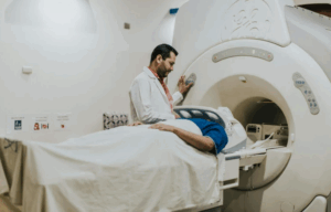
Magnetic Resonance Imaging
Principle: MRI makes use of magnetic fields and radio waves in producing cross-sectional images of the body with excellence, especially on soft tissues and bones.
Procedure:
The patient is placed inside a cylindrical magnet, on a table that glides in.
Radio waves cause the misalignment of hydrogen atoms in the body due to the magnetic field.
The atoms return to their normal position when the radio waves are turned off, emitting signals that are detected and used to create images.
Application
Imaging of bone marrow disorders
Assessment of joints structures
Diagnosis of musculoskeletal tumors
Bone Scintigraphy (Bone scan)
Principle: Bone scintigraphy is performed where a small amount of radioactive tracer is injected intravenously and collects in bones with high metabolic activity.
Procedure:
It is administered intravenously by injecting a radiotracer into the patient’s body.
After a waiting period, the patient would lie on a table, and then a gamma camera will pick up the radiation emitted by the radiotracer.
In the image, areas of high radiotracer uptake will appear, where there is a more significant quantity of bone activity.
Applications
Bone metastases detection
Diagnosis of the fracture which doesn’t appear in the X-ray films
Stage of the bone infection
Dual-energy X-ray Absorptiometry (DEXA)
Principle: DEXA utilises a low intensity low energy X-ray beam and a high intensity high energy X ray beam to determine the bone mineral content.
Procedure:
This can be done at bedside of patient in which patient lies on table and X-ray generator passed over body.
X-ray tubes emit X-ray beams that pass-through bones and the intensity of X-rays absorbed by bones is measured.
A computer calculates bone mineral density (BMD).
Applications
Diagnosing osteoporosis
Assessing fracture risk
Laboratory tests
X-rays
It uses electromagnetic radiation to make the bones and other structures inside the body visible on a screen.
Computed Tomography (CT) Scan
CT scans are images produced using X-rays and computer technology to display cross-sectional images of the body.
Magnetic Resonance Imaging (MRI)
MRI entails the use of powerful magnets and radio waves in formation of remarkable images of bones, joints and the soft tissues.
Bone Scintigraphy (Bone Scan)
A Bone scan entails the administration of a small tracer of radioactive substance into the body and then takes pictures of the bone with a gamma camera.
(DEXA) Scan
DEXA scans are low-energy X-ray scans for the assessment of bone mineral density.
Ultrasound
It uses sound waves of high frequencies to devise pictures of soft tissue, muscles, and joint areas.
PET scan
PET stands for Positron Emission Tomography.
PET scans are imaging techniques in which a radioactive tracer is utilized to help detect disease within the body.
Complications
Radiation Exposure:
X-rays and CT scans: It uses ionizing radiation and therefore there is high potential for cancer development particularly in long term usage with many practices.
Allergic Reactions:
Contrast Agents: Utilised in CT scans and MRI to improve image quality. Some patients may have allergy to this product that may range from mild to severe such as rash, itching to anaphylactic reactions.
Kidney Damage:
Contrast-Induced Nephropathy: Contrast agents are toxic to the kidneys especially to patients with impaired kidney function.
Metal Interference:
MRIs: Some of other precaution that need to be taken is that patient with implanted material such as pacemaker, cochlear implants or metal clips might be in danger since the magnetic field can affect the function of such implants.
Claustrophobia and Anxiety:
MRIs: Some people may find MRIs uncomfortable due to the confined space of the machine, or they may even develop claustrophobia.
Diagnostic Errors:
Images might be misread; therefore, incorrect diagnosis might occur.
Musculoskeletal radiology or skeletal imaging involves imaging of the skeleton and its other, internally related, body structures. It is used in the diagnosis and treatment of innumerable disorders that affect the skeletal framework, such as fractures, infections, arthritis, tumors, and congenital anomalies.
Fracture Diagnosis
These methods can be used to diagnose the points of fracture and, according to their nature, their assessment can be made as to whether they are severe or not.
Diagnosis of stress fracture that may not show in regular X-ray tests.
Bone and Joint Diseases:
Diagnosis of the potential for arthritis and evaluation of how severe and what type of arthritis it is.
Diagnosis of bone infections (osteomyelitis).
Tumor Detection:
Diagnosis of primary bone tumors or other tumors which metastasize to the bone.
Congenital and Developmental Disorders:
Evaluation of congenital skeletal anomalies.
Diagnostic testing of developmental dysplasia of the hip in infants.
Trauma:
Measuring tissue damage from injury like a car accident, a slip, or a fall.
X-rays and CT scans:
Pregnancy: Because of the possibility of the effects that the ionizing radiation can have on the fetus.
Allergy to Contrast Media: In the case of contrast-enhanced CT scans.
Severe Renal Impairment: Caused by the opposition of contrast agents on kidney tissues and the possibility of contrast-induced nephropathy.
Unstable Patients: Mobile patients, which means patients unable to stay in one position or those in a very poor health condition.
MRI Scans:
Metal Implants or Devices: Those with certain types of metal implants in their body which are not MRI safe such as pacemakers, cochlear implants and some aneurysm clips.
Allergy to Gadolinium: This is used for MRI scans when contrast agents are used.
Bone Scans:
Pregnancy and Breastfeeding: As a result of the application of radioactive tracers.
X-ray Machines
Computed Tomography (CT) Scanners
Magnetic Resonance Imaging (MRI) Machines
Bone Densitometers (DEXA Scanners)
Fluoroscopy Machines
Ultrasound Machines
Nuclear Medicine Equipment
Positron Emission Tomography (PET) Scanners
Clothes and Accessories:
Patients should wear comfortable clothes that are not going to interfere with the machinery; this includes clothes not having zippers or metal buttons.
Patients may need to wear hospital gowns in some cases.
Jewellery and Metal Objects:
All objects that can include jewellery, watches, eyeglasses and all metals are to be removed since they have an influence on the images to be produced.
Medical History:
A patient should disclose any disorder, disease or operation they have had within the last six months with the technologist.
Female patients must notify if there is any possibility of pregnancy.
Fasting:
There are cases when the patient is asked to fast before the intervention, depending on the type of imaging.
The patient will be positioned in a way depending on the area that needs imaging as determined by the physician.
Technologists will ensure the patient is comfortable and properly aligned.
Principle: X-rays are a type of electromagnetic radiation which being penetrated in the body can provide images of the internal structures of the body.
Procedure:
A patient stands facing the X-ray source with the detector on the opposite side of them.
This is the case as X-rays are taken from the source and penetrate the human body.
X-rays penetrate through tissues and bones in different manners; bones are dense and allow few X-rays to pass through them and hence appear white while soft tissues, being less dense, allow more X-rays to get through them and so appear darker on the radiograph.
Applications
Detecting fractures
Identifying bone infections
As for the evaluation of joint disorders such as arthritis.
Principle: CT uses X-rays to create detailed cross-sectional images of the body by rotating around the patient and using computer processing to create a series of slices.
Procedure:
Person receiving the CT scans lie on a table in a circular path or ‘gantry’ of the CT scanner.
It remains mounted above the patient, and the patient is moved, or the X-ray tube is revolved to capture the image from several directions.
The obtained images are then analyzed by a computer to formulate accurate cross-sectional images.
Applications
There was an evidenced-based conclusion on the assessment of multi-faceted fractures.
Assessment of bone tumors
Planning orthopedic surgeries
Principle: MRI makes use of magnetic fields and radio waves in producing cross-sectional images of the body with excellence, especially on soft tissues and bones.
Procedure:
The patient is placed inside a cylindrical magnet, on a table that glides in.
Radio waves cause the misalignment of hydrogen atoms in the body due to the magnetic field.
The atoms return to their normal position when the radio waves are turned off, emitting signals that are detected and used to create images.
Application
Imaging of bone marrow disorders
Assessment of joints structures
Diagnosis of musculoskeletal tumors
Bone Scintigraphy (Bone scan)
Principle: Bone scintigraphy is performed where a small amount of radioactive tracer is injected intravenously and collects in bones with high metabolic activity.
Procedure:
It is administered intravenously by injecting a radiotracer into the patient’s body.
After a waiting period, the patient would lie on a table, and then a gamma camera will pick up the radiation emitted by the radiotracer.
In the image, areas of high radiotracer uptake will appear, where there is a more significant quantity of bone activity.
Applications
Bone metastases detection
Diagnosis of the fracture which doesn’t appear in the X-ray films
Stage of the bone infection
Principle: DEXA utilises a low intensity low energy X-ray beam and a high intensity high energy X ray beam to determine the bone mineral content.
Procedure:
This can be done at bedside of patient in which patient lies on table and X-ray generator passed over body.
X-ray tubes emit X-ray beams that pass-through bones and the intensity of X-rays absorbed by bones is measured.
A computer calculates bone mineral density (BMD).
Applications
Diagnosing osteoporosis
Assessing fracture risk
X-rays
It uses electromagnetic radiation to make the bones and other structures inside the body visible on a screen.
Computed Tomography (CT) Scan
CT scans are images produced using X-rays and computer technology to display cross-sectional images of the body.
Magnetic Resonance Imaging (MRI)
MRI entails the use of powerful magnets and radio waves in formation of remarkable images of bones, joints and the soft tissues.
Bone Scintigraphy (Bone Scan)
A Bone scan entails the administration of a small tracer of radioactive substance into the body and then takes pictures of the bone with a gamma camera.
(DEXA) Scan
DEXA scans are low-energy X-ray scans for the assessment of bone mineral density.
Ultrasound
It uses sound waves of high frequencies to devise pictures of soft tissue, muscles, and joint areas.
PET scan
PET stands for Positron Emission Tomography.
PET scans are imaging techniques in which a radioactive tracer is utilized to help detect disease within the body.
Radiation Exposure:
X-rays and CT scans: It uses ionizing radiation and therefore there is high potential for cancer development particularly in long term usage with many practices.
Allergic Reactions:
Contrast Agents: Utilised in CT scans and MRI to improve image quality. Some patients may have allergy to this product that may range from mild to severe such as rash, itching to anaphylactic reactions.
Kidney Damage:
Contrast-Induced Nephropathy: Contrast agents are toxic to the kidneys especially to patients with impaired kidney function.
Metal Interference:
MRIs: Some of other precaution that need to be taken is that patient with implanted material such as pacemaker, cochlear implants or metal clips might be in danger since the magnetic field can affect the function of such implants.
Claustrophobia and Anxiety:
MRIs: Some people may find MRIs uncomfortable due to the confined space of the machine, or they may even develop claustrophobia.
Diagnostic Errors:
Images might be misread; therefore, incorrect diagnosis might occur.
Both our subscription plans include Free CME/CPD AMA PRA Category 1 credits.
Digital Certificate PDF
On course completion, you will receive a full-sized presentation quality digital certificate.
medtigo Simulation
A dynamic medical simulation platform designed to train healthcare professionals and students to effectively run code situations through an immersive hands-on experience in a live, interactive 3D environment.
medtigo Points
medtigo points is our unique point redemption system created to award users for interacting on our site. These points can be redeemed for special discounts on the medtigo marketplace as well as towards the membership cost itself.
Community Forum post/reply = 5 points
*Redemption of points can occur only through the medtigo marketplace, courses, or simulation system. Money will not be credited to your bank account. 10 points = $1.
All Your Certificates in One Place
When you have your licenses, certificates and CMEs in one place, it's easier to track your career growth. You can easily share these with hospitals as well, using your medtigo app.