Skin and Soft Tissue Infections – Incision, Drainage and Debridement
Updated :
December 29, 2025
Background
Skin and soft tissue infections (SSTIs) are infections that affect skin, subcutaneous tissue, fascia and muscle and are caused by various pathogenic microorganisms such as Staphylococcus aureus and Streptococcus pyogenes or site-specific infections or immunocompromised hosts, monomicrobial necrotizing fasciitis or polymicrobial necrotizing fasciitis.
Simple soft tissue infections (SSTIs) consist of superficial cellulitis, folliculitis, furunculosis, simple abscesses, and small wound infections. These can be treated by either addressing the cause or using antibiotics. Hence, they have relatively low mortality rates or chances of leading to amputation. On the other hand, complicated SSTIs are characterized by invasion into deeper structures hence need serious surgical intervention most times, an issue which may further complicate drug treatment due to comorbidities present in such cases.
Necrotising fasciitis is an increasingly aggressive, fast-moving and inflammatory infection found in the deep fascia with secondary necrosis of the hypodermis. It is this depth that causes thrombosis in skin vessels leading to death of the epidermis due to lack of blood supply through them.
It is important to distinguish between necrotizing infections and those that do not necrotize, if one is to manage them successfully. There are various factors that make people more likely to develop necrotizing soft tissue infections that includes skin injury, dry irritated skin, being immunocompromised due to any reason, having chronic venous insufficiency, chronic lymphatic insufficiency, or some form of nerve damage that are old and not new. Saving life is possible with timely diagnosis and treatment, otherwise an affected person can lose his/her leg or even die.
Indications
Every abscess, no matter how small, has tobe washed out completely. Dead and necrotic tissues covering any ulcer must be removed so that healthy granulation tissue growth and healing can take place optimally. Immediate surgical intervention is necessary for necrotizing fasciitis, which when carried out early, the surgery results in the best outcomes in such patients.
Contraindications
Thereare no absolute contraindications for these procedures. If the physical condition of the patient is compromised, stabilization to render him or her fit for anesthesia should be carried out before these procedures are used.
Outcomes
It is important to have the right antibiotic treatment in difficult to treat skin infections. Identifying and treating this kind of an infection quickly is crucial because prolonging the period before beginning medical care leads to significant rates as far as diseases seriousness and deaths.
Equipment
A basic surgical set is the equipment necessary for the procedure of incision, drainage and debridement. This includes the following:
Nos 11 and 15 surgical blades
Curette
Sponge-holding forceps
Metzenbaum scissors
Electrocautery
Curved artery forceps
Sterile swab stick
Toothed and plane thumb forceps
Hydrogen peroxide
Saline
Patient preparation
Adequate anesthesia and correct positioning are part of patient preparation.
Anesthesia
Anesthesia for incision and drainage includes general anesthesia for large and deep abscesses, regional anesthesia for large and deep abscesses, and field block for small to medium-sized abscesses. Local anesthesia is used for painless incisions but is less effective in the acidic environment of the abscess cavity. General anesthesia is preferred for debridement, as it is painful and requires complete analgesia for thorough debridement. Regional anesthesia or field block can be used when general anesthesia is not desirable and patient cooperation is ensured. Field block is suitable for small to medium-sized abscesses.
Anesthesia for debridement complies of general or field block anesthesia as the procedure is very painful.
Patient position
The patient is positioned depending on the location of the problem. The surgeon should be able to get to the problem area easily and the patient should not feel uncomfortable throughout the procedure.
Approach considerations
When draining an abscess, make an incision at the point of maximal fluctuance, ideally in a dependent area, and align with the crease of natural skin. Bold incision must be used to reach the abscess cavity, and if major vessels or nerves are present, blunt dissection should be performed. Excision of necrosed or unhealthy skin on the roof of the abscess is crucial. Debridement should be continued until healthy dermal bleeding is noticed at the edges of the skin with no loose, undermined skin edges. Avoid overzealous debridement in non-necrotizing skin and soft tissue infections (SSTIs) and plan staged debridement to minimize damage to healthy tissue.
Technical considerations
Step-1: Incision and drainage
In the process of incision and drainage of an abscess, a skin incision should be made parallel to the natural skin folds in order to minimize scarring. A bold incision can be made through the skin, subcutaneous tissue and deep fascia where there are no significant neural or vascular structures. In places where there are significant neurovascular structures, however, only the skin and subcutaneous tissue are incised while the abscess cavity is accessed by gently inserting blunt artery forceps or sinus forceps through a small opening in the deep fascia. As soon as pus starts extruding from the opening, the cavity should be entered and explored with artery forceps armed with a gauze piece or the surgeon’s gloved finger. Once the cavity has been entered, it should be washed out with normal saline solution and packed with gauze or a surgical sponge.
Step-2: Debridement for gangrene and infected ulcers
Debridement is a surgical procedure that involves cleaning and draping around the lesion area, carefully excising the slough from the underlying healthy tissue, and freshening the wound margins. The tourniquet should then be released, and any active points of bleeding should be cauterised. Non-adherent dressing like tulle grars should be used to cover raw areas and changed for every 24 hours. The wound should also be checked for new slough or pus formation at every change of dressing. It may be necessary to have several debridements until the wound is clean and the control of infection with antibiotics. When wound is clean, dressings can be changed every 36 hours.
Other methods of debridement other than surgical debridement include:
Chemical
Autolytic
Mechanical
Ultrasonic
Biological
Mechanical debridement involves the application of a moist dressing followed by a dry one that removes adherent tissue. Enzymatic agents are used in chemical debridement to dissolve dead tissue before regular dressings are applied. Autolytic debridement utilizes the body’s own moisture and matrix metalloproteinases (MMPs) to help slough off necrotic tissue. Dressings that maintain a moist environment retards this process. Biological debridement (maggot therapy) is achieved through the use of greenbottle fly maggots, which feed on necrotic tissue and bacteria while sparing living tissue. Ultrasonic debridement causes erosion and disruption of necrotic tissues through cavitation created by ultrasonic vibrations leading to tissue fragmentation and emulsification. However, patients are generally reluctant to accept this method because its safety remains questionable.
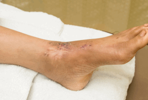
Debridement on ankle
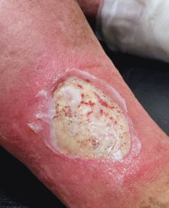
Wound Debridement
Step-3: Debridement for necrotizing fasciitis
In diagnosing necrotizing fasciitis, a tourniquet is used to create a bloodless field. All three zones of fasciitis are marked off, and then along with a bold incision extending the length of the lesion. By probing its limits, we can ascertain the extent of the deep fascia. Tissue samples for culture, antibiotic sensitivity testing, and histopathological examination should be taken. After cutting out the deep fascia up to healthy muscle underneath exposed by radical excision, check skin overlying them together with subcutaneous tissues viability should be done. Zone 1 tissues need to be excised while zone 2 ones must only undergo general observation regarding their viability. The tourniquet gets released so that all tissues can get rechecked for viability including deflated ones. Haemostasis achieved using electrocautery then wound thoroughly washed with saline. Non-adherent dressing must be applied over wound and then covered by absorbent bandage, which is changed after 24 hours.
Laboratory tests
Patients with uncomplicated SSTIs usually do not require hospitalization or investigations. However, immunocompromised patients or those exhibiting signs and symptoms of systemic toxicity such as hypotension and tachycardia may need the following tests:
Wound culture
Creatinine levels
CBC (complete blood count) with differential
Bicarbonate level
Level of C-reactive protein
Drug susceptibility and blood culture
Complications
Bleeding is a common complication in surgical procedures, especially in cases of necrotizing fasciitis, which can lead to rapid deterioration of the patient. Proper hemostasis is crucial, and pressure dressings are usually sufficient. Severe sepsis can occur from the dissemination of bacteria and toxins into the bloodstream, so it is essential to perform these procedures under treatment with antibiotics. Additionally, underlying nerves and blood vessels can be injured, so surgeons must be cautious during incisions and debridement, especially if the abscess or ulcer is near vital anatomical structures.
»
Home » Procedure » Skin and Soft Tissue Infections – Incision, Drainage and Debridement
Skin and Soft Tissue Infections – Incision, Drainage and Debridement
Updated :
December 29, 2025
Skin and soft tissue infections (SSTIs) are infections that affect skin, subcutaneous tissue, fascia and muscle and are caused by various pathogenic microorganisms such as Staphylococcus aureus and Streptococcus pyogenes or site-specific infections or immunocompromised hosts, monomicrobial necrotizing fasciitis or polymicrobial necrotizing fasciitis.
Simple soft tissue infections (SSTIs) consist of superficial cellulitis, folliculitis, furunculosis, simple abscesses, and small wound infections. These can be treated by either addressing the cause or using antibiotics. Hence, they have relatively low mortality rates or chances of leading to amputation. On the other hand, complicated SSTIs are characterized by invasion into deeper structures hence need serious surgical intervention most times, an issue which may further complicate drug treatment due to comorbidities present in such cases.
Necrotising fasciitis is an increasingly aggressive, fast-moving and inflammatory infection found in the deep fascia with secondary necrosis of the hypodermis. It is this depth that causes thrombosis in skin vessels leading to death of the epidermis due to lack of blood supply through them.
It is important to distinguish between necrotizing infections and those that do not necrotize, if one is to manage them successfully. There are various factors that make people more likely to develop necrotizing soft tissue infections that includes skin injury, dry irritated skin, being immunocompromised due to any reason, having chronic venous insufficiency, chronic lymphatic insufficiency, or some form of nerve damage that are old and not new. Saving life is possible with timely diagnosis and treatment, otherwise an affected person can lose his/her leg or even die.
Every abscess, no matter how small, has tobe washed out completely. Dead and necrotic tissues covering any ulcer must be removed so that healthy granulation tissue growth and healing can take place optimally. Immediate surgical intervention is necessary for necrotizing fasciitis, which when carried out early, the surgery results in the best outcomes in such patients.
Thereare no absolute contraindications for these procedures. If the physical condition of the patient is compromised, stabilization to render him or her fit for anesthesia should be carried out before these procedures are used.
It is important to have the right antibiotic treatment in difficult to treat skin infections. Identifying and treating this kind of an infection quickly is crucial because prolonging the period before beginning medical care leads to significant rates as far as diseases seriousness and deaths.
A basic surgical set is the equipment necessary for the procedure of incision, drainage and debridement. This includes the following:
Nos 11 and 15 surgical blades
Curette
Sponge-holding forceps
Metzenbaum scissors
Electrocautery
Curved artery forceps
Sterile swab stick
Toothed and plane thumb forceps
Hydrogen peroxide
Saline
Adequate anesthesia and correct positioning are part of patient preparation.
Anesthesia
Anesthesia for incision and drainage includes general anesthesia for large and deep abscesses, regional anesthesia for large and deep abscesses, and field block for small to medium-sized abscesses. Local anesthesia is used for painless incisions but is less effective in the acidic environment of the abscess cavity. General anesthesia is preferred for debridement, as it is painful and requires complete analgesia for thorough debridement. Regional anesthesia or field block can be used when general anesthesia is not desirable and patient cooperation is ensured. Field block is suitable for small to medium-sized abscesses.
Anesthesia for debridement complies of general or field block anesthesia as the procedure is very painful.
Patient position
The patient is positioned depending on the location of the problem. The surgeon should be able to get to the problem area easily and the patient should not feel uncomfortable throughout the procedure.
When draining an abscess, make an incision at the point of maximal fluctuance, ideally in a dependent area, and align with the crease of natural skin. Bold incision must be used to reach the abscess cavity, and if major vessels or nerves are present, blunt dissection should be performed. Excision of necrosed or unhealthy skin on the roof of the abscess is crucial. Debridement should be continued until healthy dermal bleeding is noticed at the edges of the skin with no loose, undermined skin edges. Avoid overzealous debridement in non-necrotizing skin and soft tissue infections (SSTIs) and plan staged debridement to minimize damage to healthy tissue.
Step-1: Incision and drainage
In the process of incision and drainage of an abscess, a skin incision should be made parallel to the natural skin folds in order to minimize scarring. A bold incision can be made through the skin, subcutaneous tissue and deep fascia where there are no significant neural or vascular structures. In places where there are significant neurovascular structures, however, only the skin and subcutaneous tissue are incised while the abscess cavity is accessed by gently inserting blunt artery forceps or sinus forceps through a small opening in the deep fascia. As soon as pus starts extruding from the opening, the cavity should be entered and explored with artery forceps armed with a gauze piece or the surgeon’s gloved finger. Once the cavity has been entered, it should be washed out with normal saline solution and packed with gauze or a surgical sponge.
Step-2: Debridement for gangrene and infected ulcers
Debridement is a surgical procedure that involves cleaning and draping around the lesion area, carefully excising the slough from the underlying healthy tissue, and freshening the wound margins. The tourniquet should then be released, and any active points of bleeding should be cauterised. Non-adherent dressing like tulle grars should be used to cover raw areas and changed for every 24 hours. The wound should also be checked for new slough or pus formation at every change of dressing. It may be necessary to have several debridements until the wound is clean and the control of infection with antibiotics. When wound is clean, dressings can be changed every 36 hours.
Other methods of debridement other than surgical debridement include:
Chemical
Autolytic
Mechanical
Ultrasonic
Biological
Mechanical debridement involves the application of a moist dressing followed by a dry one that removes adherent tissue. Enzymatic agents are used in chemical debridement to dissolve dead tissue before regular dressings are applied. Autolytic debridement utilizes the body’s own moisture and matrix metalloproteinases (MMPs) to help slough off necrotic tissue. Dressings that maintain a moist environment retards this process. Biological debridement (maggot therapy) is achieved through the use of greenbottle fly maggots, which feed on necrotic tissue and bacteria while sparing living tissue. Ultrasonic debridement causes erosion and disruption of necrotic tissues through cavitation created by ultrasonic vibrations leading to tissue fragmentation and emulsification. However, patients are generally reluctant to accept this method because its safety remains questionable.
Debridement on ankle
Wound Debridement
Step-3: Debridement for necrotizing fasciitis
In diagnosing necrotizing fasciitis, a tourniquet is used to create a bloodless field. All three zones of fasciitis are marked off, and then along with a bold incision extending the length of the lesion. By probing its limits, we can ascertain the extent of the deep fascia. Tissue samples for culture, antibiotic sensitivity testing, and histopathological examination should be taken. After cutting out the deep fascia up to healthy muscle underneath exposed by radical excision, check skin overlying them together with subcutaneous tissues viability should be done. Zone 1 tissues need to be excised while zone 2 ones must only undergo general observation regarding their viability. The tourniquet gets released so that all tissues can get rechecked for viability including deflated ones. Haemostasis achieved using electrocautery then wound thoroughly washed with saline. Non-adherent dressing must be applied over wound and then covered by absorbent bandage, which is changed after 24 hours.
Laboratory tests
Patients with uncomplicated SSTIs usually do not require hospitalization or investigations. However, immunocompromised patients or those exhibiting signs and symptoms of systemic toxicity such as hypotension and tachycardia may need the following tests:
Wound culture
Creatinine levels
CBC (complete blood count) with differential
Bicarbonate level
Level of C-reactive protein
Drug susceptibility and blood culture
Complications
Bleeding is a common complication in surgical procedures, especially in cases of necrotizing fasciitis, which can lead to rapid deterioration of the patient. Proper hemostasis is crucial, and pressure dressings are usually sufficient. Severe sepsis can occur from the dissemination of bacteria and toxins into the bloodstream, so it is essential to perform these procedures under treatment with antibiotics. Additionally, underlying nerves and blood vessels can be injured, so surgeons must be cautious during incisions and debridement, especially if the abscess or ulcer is near vital anatomical structures.
Both our subscription plans include Free CME/CPD AMA PRA Category 1 credits.
Digital Certificate PDF
On course completion, you will receive a full-sized presentation quality digital certificate.
medtigo Simulation
A dynamic medical simulation platform designed to train healthcare professionals and students to effectively run code situations through an immersive hands-on experience in a live, interactive 3D environment.
medtigo Points
medtigo points is our unique point redemption system created to award users for interacting on our site. These points can be redeemed for special discounts on the medtigo marketplace as well as towards the membership cost itself.
Community Forum post/reply = 5 points
*Redemption of points can occur only through the medtigo marketplace, courses, or simulation system. Money will not be credited to your bank account. 10 points = $1.
All Your Certificates in One Place
When you have your licenses, certificates and CMEs in one place, it's easier to track your career growth. You can easily share these with hospitals as well, using your medtigo app.