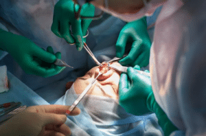
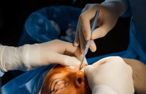
Reconstruction needs a strong knowledge of how the skin is structured, diagnosis of the defects, and the selection of the best donor site. A “ladder” is used to classify the options and these include secondary intention healing, pedicle, and random flaps, and microvascular, free tissue transfer. For local flaps modifications are least time consuming option with a reliable blood supply and similar color / texture of skin.
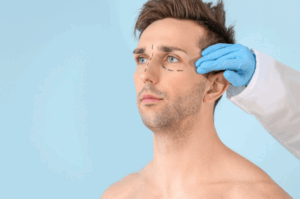
The subunit principle is the key to proper analysis of facial defects and determination of the most suitable way to conceal the incision borders and camouflage transitions by considering options such as skin color, texture, thickness, hair growth, as well as the facial contours.
The flap choice depends on good match, minimal distortion, and adequate area of reconstruction. Plastic anaesthetic surgeons must hold the local flaps in order to visualize and estimate consequent success.
Facial subunits
Indications
Contraindications
Outcomes
Local Flaps
The microcirculatory system has a vascular deep plexus and superficial plexus which transport blood flow to epidermis in a diffusive and flow in collaterals. This microcirculation is supported by septocutaneous and musculocutaneous arteries.
The skin is innervated by both sensory and sympathetic nervous systems; sensory afferents are distributed territorially, while arteriaiolar preshunt sphincters of those vessel are controlled by sympathetic afferents. Cold-water split at flap edges dissects plexus layers resulting in vascular resistance unrendering norepinephrine and blocks catecholamine reuptake.
Blood supply to the flap base is an essential condition for the success of the flap. Besides the formation of new vascular channels between the flap and the recipient bed is also critical. The perfusion pressure is the major determinant of flap length, where a base length adjustment of only 50% may not result in different flap length.
A neovascularization of a flap develops for a period of 3 to 7 days after the transfer of the flap into nonvascularized area through direct ingrowth processes and inosculation. A vasodilator released from the flap edges in turn causes an angiogenic reaction by promoting migration of endothelial cells and capillary sprouts’ formation.
Classification of Flaps
Flaps can be grouped into two main groups based on the blood supply construction, geometry, anatomy, and transposition methods.Itbreaks down into five methods but these are largely combinations of these classification which is based on the advancement, pivotal, transpositional, rhombic, and bilobed flaps.
Advancement flaps
Advancement flaps are linear or rectangular, subclasses include simple, single pedicle, bipedicle, and V-Y flaps.
Single-pedicle advancement flaps
Parallel incisions are done on flaps locate at the site of the desresct repurposed as a donor site, advanced into the defect, diminishing tension and healing it with a nice line of scar. Cone deformities can be taken out through three ways.
This principle of halves, Burow’s triangles, and bilateral z-plasties gives flapwidth enough to allow overlong flaps to gain necessary length and rid them of the standing cone.
The single-pedicle dermal flaps (island advancement) are a special flap designed to close the defect by xanthumum nigra medium (the dermis through which incised to preserve sub dermis tissue), and closed in a V-Y fashion.
Bipedicle advancement flaps
Bipedicle local flaps are an alternative perpendicular to the defect axis, which are used to remove defects under the high visibility areas and then move them to low visibility areas but are less common due to easiest and more aesthetic reconstruction option.
The V-Y advancement flap
The advancement flap has a uniqueness in which the donor defect is being pushed with a flap that is made in such a way that it would resemble in a straight line the suture and it’s leading edge would be shaped like a wound’s edge this will create a scar shaped like a lollipop.
Pivotal flaps
Pivotal flaps, which are rotation, transposition, and interposition flaps, are used to go from the donor site to the defective site.
Rotation flaps
The rotational flap method is used for defects with curvilinear edges that are perpendicular to the direction of rotation. This improves lymphatic drain and gives the perfect cover. The flaps must be longer than the defect’s border by a 4:1 ratio.
Transposition flaps
Transposition flaps are multifunctional flaps are utilized for head and neck defect repair giving the option of compatible colors and texture from different donor sites. They require a 3:1 vascularity in the head and neck helps for flaps exceeding the standard 1:1 length-to-width ratio.
Z-plasty is a facial reconstruction technique that utilizes flaps that are rotated in the same angles, stretching the contracted scars, changing the direction of the scars, and cutting the linearity.
Interpolation flaps
Interpolation flap resembling transposition flap shifts around pedicle and transposes over intervening tissue with the resting place. Pedicle divided and inset after vascularization is done, common forehead flap. Rhombic Flaps Limberg devised the rhombic flap, a transposition flap intended to treat a rhomboid defect with sides of equal length and a short diagonal line. The rhombic defects have 4 closure flaps. To establish the best flap, the lines of maximum extensibility (LMEs) are drawn around the defect. We have two lines on the same plane, with 60° and 120° angles. The short unit is replicated, forming two flaps. The surgeon needs to decide the precise flap that will be the best match to the skin and hide the scar. The Dufourmentel flap is an alternative to the classic Limberg rhombic flap, which only works with defects of the shape rhomb with two opposite corners of 60° and 120°. Bilobed Flaps The bilobed flap design is a second type of transposition flap which consists of two transposition flaps with a 90° angle between them. It is used to repair defects and donor sites, but may cause standing cone deformities and pincushioning. A modification reduces angles to 45°. The design of a bilobed flap takes into account tissue elasticity, possible deformation, and RSTLs; often with the use of dermabrasion to reduce scarring. It is good with ala and tip defects in the nose.
Reconstruction of Facial Subunits
The complicated procedure of picking out the more effective artificial method for a specific cut, one which focuses on the tissue flaps and their pitfalls. we must always draw the peripheral hubs and close the flap roof before stitching. This is to minimize tension and ease the closing. Nose Nose is the part that appears most often in trauma and burns. If the 3-layered technique is appropriately carried out, function is preserved and severe cramping is avoided. In addition, the surgery may lead to scar contraction especially at the nasal alar subunit. This situation can result in deformity or even airway obstruction. The first two thirds of the nose that are found to be defective are often repaired with thinner skin that has less oil glands. The paramedian forehead skin flap, an interpolation flap which is- enriched with an abundant amount of tissue as well as matches successfully the color and texture of the patient’s original skin, offers an excellent opportunity for resurfacing the entire surface of the patient’s nose. The spatial position of alar demands cartilage means to prevent upward contracture. One of the common donor sites that is utilized for the external lining is the melolabial or the paramedian forehead flap, for defects that are adjacent to the lateral subunit. Reconstructive difficulties like scarring and expedient address through Z-plasty and cheek advancement flaps.
Nose reconstruction
Cheek In cases of cheek corrections the depth variations and curves of different sides of the cheek must be considered. Primary closure is better for minor defects hence RSTLs are preferred the most and for the medium-to-large one’s local flap coverage is needed. The rhombic and transposition flap types help to shape the cheeks. The incisions should, however, not be perpendicular to the RSTLs, that are limited in lateral and upper cheek. Tissue advancement rotation flaps that are used in cheek reconstruction and allow recruitment of tissues without any secondary deformities are often the local flaps of choice in that region. Underside of the incision should be medial, lateral to the orbital rim and infraorbital margin may be repaired by island pedicle flaps.
Lip
Lip reconstruction aims at sustaining oral competency, motor, and sensory supply, and providing a sulcus that will not be distorted. The first step is not only considering the entire sphincter, but also, if the upper lip is not attainable, giving priority to the lower lip. Red lip reconstruction is done with tissue from either red lip or buccal mucosa and small defects are fixed with V-Y advancement flaps or Z-plasty while more extensive defects may require vermilionectomy and a buccal mucosal flap. For small lip scars, excisions with primary closure should be carried out in order to avoid notching and the usage of flap extensions. In some cases, deeper defects can be reconstructed by using inferiorly based melolabial flaps, but this might erase the melolabial crease. Secondary fold creation can become the beauty that one seeks after. Eyelid In eyelid reconstruction, the object is to restore eyelids that are functional and include both anterior and posterior lamellae. This needs to be used for full-thickness defects, so as to protect the globe and provide appropriate cosmetics. Closure of partial-thickness lower lid defects which cannot be closed can be done by advancement rotation flaps or by transposing upper lid to lower lid flaps, and thicker or thinner skin defects are usually reconstructed by using grafts.
Eyelid reconstruction
Forehead In general, forehead pattern reconstruction is achieved byadvancement and rotation flaps, except when a vertical scar crosses RSTL at a right angle for cosmetic approval. The uncapped bellies of frontalis muscles decrease the effect in the midline and paramedian regions of the forehead’s vertical movement, minimizing vertical scar distortion. H-plasty is the most popular type of forehead defect closure using bilateral advancement flaps, variations include O-T, A-T, and O-Z closures applied in similar reconstructions. Scalp The rotation advancement flap is a well-known method of scalp reconstruction for central or vertex defects of 3-4 cm. The Orticochea flap, a modification of the 3-flap technique, is chosen for larger defects, able to provide 10-12 cm of closure.
Reconstruction needs a strong knowledge of how the skin is structured, diagnosis of the defects, and the selection of the best donor site. A “ladder” is used to classify the options and these include secondary intention healing, pedicle, and random flaps, and microvascular, free tissue transfer. For local flaps modifications are least time consuming option with a reliable blood supply and similar color / texture of skin.
The subunit principle is the key to proper analysis of facial defects and determination of the most suitable way to conceal the incision borders and camouflage transitions by considering options such as skin color, texture, thickness, hair growth, as well as the facial contours.
The flap choice depends on good match, minimal distortion, and adequate area of reconstruction. Plastic anaesthetic surgeons must hold the local flaps in order to visualize and estimate consequent success.
Facial subunits
The microcirculatory system has a vascular deep plexus and superficial plexus which transport blood flow to epidermis in a diffusive and flow in collaterals. This microcirculation is supported by septocutaneous and musculocutaneous arteries.
The skin is innervated by both sensory and sympathetic nervous systems; sensory afferents are distributed territorially, while arteriaiolar preshunt sphincters of those vessel are controlled by sympathetic afferents. Cold-water split at flap edges dissects plexus layers resulting in vascular resistance unrendering norepinephrine and blocks catecholamine reuptake.
Blood supply to the flap base is an essential condition for the success of the flap. Besides the formation of new vascular channels between the flap and the recipient bed is also critical. The perfusion pressure is the major determinant of flap length, where a base length adjustment of only 50% may not result in different flap length.
A neovascularization of a flap develops for a period of 3 to 7 days after the transfer of the flap into nonvascularized area through direct ingrowth processes and inosculation. A vasodilator released from the flap edges in turn causes an angiogenic reaction by promoting migration of endothelial cells and capillary sprouts’ formation.
Flaps can be grouped into two main groups based on the blood supply construction, geometry, anatomy, and transposition methods.Itbreaks down into five methods but these are largely combinations of these classification which is based on the advancement, pivotal, transpositional, rhombic, and bilobed flaps.
Advancement flaps are linear or rectangular, subclasses include simple, single pedicle, bipedicle, and V-Y flaps.
Single-pedicle advancement flaps
Parallel incisions are done on flaps locate at the site of the desresct repurposed as a donor site, advanced into the defect, diminishing tension and healing it with a nice line of scar. Cone deformities can be taken out through three ways.
This principle of halves, Burow’s triangles, and bilateral z-plasties gives flapwidth enough to allow overlong flaps to gain necessary length and rid them of the standing cone.
The single-pedicle dermal flaps (island advancement) are a special flap designed to close the defect by xanthumum nigra medium (the dermis through which incised to preserve sub dermis tissue), and closed in a V-Y fashion.
Bipedicle advancement flaps
Bipedicle local flaps are an alternative perpendicular to the defect axis, which are used to remove defects under the high visibility areas and then move them to low visibility areas but are less common due to easiest and more aesthetic reconstruction option.
The V-Y advancement flap
The advancement flap has a uniqueness in which the donor defect is being pushed with a flap that is made in such a way that it would resemble in a straight line the suture and it’s leading edge would be shaped like a wound’s edge this will create a scar shaped like a lollipop.
Pivotal flaps, which are rotation, transposition, and interposition flaps, are used to go from the donor site to the defective site.
Rotation flaps
The rotational flap method is used for defects with curvilinear edges that are perpendicular to the direction of rotation. This improves lymphatic drain and gives the perfect cover. The flaps must be longer than the defect’s border by a 4:1 ratio.
Transposition flaps
Transposition flaps are multifunctional flaps are utilized for head and neck defect repair giving the option of compatible colors and texture from different donor sites. They require a 3:1 vascularity in the head and neck helps for flaps exceeding the standard 1:1 length-to-width ratio.
Z-plasty is a facial reconstruction technique that utilizes flaps that are rotated in the same angles, stretching the contracted scars, changing the direction of the scars, and cutting the linearity.
Interpolation flaps
Interpolation flap resembling transposition flap shifts around pedicle and transposes over intervening tissue with the resting place. Pedicle divided and inset after vascularization is done, common forehead flap. Rhombic Flaps Limberg devised the rhombic flap, a transposition flap intended to treat a rhomboid defect with sides of equal length and a short diagonal line. The rhombic defects have 4 closure flaps. To establish the best flap, the lines of maximum extensibility (LMEs) are drawn around the defect. We have two lines on the same plane, with 60° and 120° angles. The short unit is replicated, forming two flaps. The surgeon needs to decide the precise flap that will be the best match to the skin and hide the scar. The Dufourmentel flap is an alternative to the classic Limberg rhombic flap, which only works with defects of the shape rhomb with two opposite corners of 60° and 120°. Bilobed Flaps The bilobed flap design is a second type of transposition flap which consists of two transposition flaps with a 90° angle between them. It is used to repair defects and donor sites, but may cause standing cone deformities and pincushioning. A modification reduces angles to 45°. The design of a bilobed flap takes into account tissue elasticity, possible deformation, and RSTLs; often with the use of dermabrasion to reduce scarring. It is good with ala and tip defects in the nose.
The complicated procedure of picking out the more effective artificial method for a specific cut, one which focuses on the tissue flaps and their pitfalls. we must always draw the peripheral hubs and close the flap roof before stitching. This is to minimize tension and ease the closing. Nose Nose is the part that appears most often in trauma and burns. If the 3-layered technique is appropriately carried out, function is preserved and severe cramping is avoided. In addition, the surgery may lead to scar contraction especially at the nasal alar subunit. This situation can result in deformity or even airway obstruction. The first two thirds of the nose that are found to be defective are often repaired with thinner skin that has less oil glands. The paramedian forehead skin flap, an interpolation flap which is- enriched with an abundant amount of tissue as well as matches successfully the color and texture of the patient’s original skin, offers an excellent opportunity for resurfacing the entire surface of the patient’s nose. The spatial position of alar demands cartilage means to prevent upward contracture. One of the common donor sites that is utilized for the external lining is the melolabial or the paramedian forehead flap, for defects that are adjacent to the lateral subunit. Reconstructive difficulties like scarring and expedient address through Z-plasty and cheek advancement flaps.
Nose reconstruction
Cheek In cases of cheek corrections the depth variations and curves of different sides of the cheek must be considered. Primary closure is better for minor defects hence RSTLs are preferred the most and for the medium-to-large one’s local flap coverage is needed. The rhombic and transposition flap types help to shape the cheeks. The incisions should, however, not be perpendicular to the RSTLs, that are limited in lateral and upper cheek. Tissue advancement rotation flaps that are used in cheek reconstruction and allow recruitment of tissues without any secondary deformities are often the local flaps of choice in that region. Underside of the incision should be medial, lateral to the orbital rim and infraorbital margin may be repaired by island pedicle flaps.
Lip
Lip reconstruction aims at sustaining oral competency, motor, and sensory supply, and providing a sulcus that will not be distorted. The first step is not only considering the entire sphincter, but also, if the upper lip is not attainable, giving priority to the lower lip. Red lip reconstruction is done with tissue from either red lip or buccal mucosa and small defects are fixed with V-Y advancement flaps or Z-plasty while more extensive defects may require vermilionectomy and a buccal mucosal flap. For small lip scars, excisions with primary closure should be carried out in order to avoid notching and the usage of flap extensions. In some cases, deeper defects can be reconstructed by using inferiorly based melolabial flaps, but this might erase the melolabial crease. Secondary fold creation can become the beauty that one seeks after. Eyelid In eyelid reconstruction, the object is to restore eyelids that are functional and include both anterior and posterior lamellae. This needs to be used for full-thickness defects, so as to protect the globe and provide appropriate cosmetics. Closure of partial-thickness lower lid defects which cannot be closed can be done by advancement rotation flaps or by transposing upper lid to lower lid flaps, and thicker or thinner skin defects are usually reconstructed by using grafts.
Eyelid reconstruction
Forehead In general, forehead pattern reconstruction is achieved byadvancement and rotation flaps, except when a vertical scar crosses RSTL at a right angle for cosmetic approval. The uncapped bellies of frontalis muscles decrease the effect in the midline and paramedian regions of the forehead’s vertical movement, minimizing vertical scar distortion. H-plasty is the most popular type of forehead defect closure using bilateral advancement flaps, variations include O-T, A-T, and O-Z closures applied in similar reconstructions. Scalp The rotation advancement flap is a well-known method of scalp reconstruction for central or vertex defects of 3-4 cm. The Orticochea flap, a modification of the 3-flap technique, is chosen for larger defects, able to provide 10-12 cm of closure.
Both our subscription plans include Free CME/CPD AMA PRA Category 1 credits.
Digital Certificate PDF
On course completion, you will receive a full-sized presentation quality digital certificate.
medtigo Simulation
A dynamic medical simulation platform designed to train healthcare professionals and students to effectively run code situations through an immersive hands-on experience in a live, interactive 3D environment.
medtigo Points
medtigo points is our unique point redemption system created to award users for interacting on our site. These points can be redeemed for special discounts on the medtigo marketplace as well as towards the membership cost itself.
Community Forum post/reply = 5 points
*Redemption of points can occur only through the medtigo marketplace, courses, or simulation system. Money will not be credited to your bank account. 10 points = $1.
All Your Certificates in One Place
When you have your licenses, certificates and CMEs in one place, it's easier to track your career growth. You can easily share these with hospitals as well, using your medtigo app.