Subtotal petrosectomy (STP) is a surgical procedure that removes air cells from the temporal bone located near the skull base.
This procedure addresses chronic infections, CSF leaks, and ensures a stable surgical field for cochlear implants.
Temporal bone surgery evolved from radical mastoidectomy for infections treatment.
The development of the operating microscope and better anatomical understanding the emergence of STP for safe surgeries.
STP is crucial in lateral skull base surgery combined with procedures such as blind sac closure, cochlear implantation, and CSF otorrhea management.
Subtotal petrosectomy effectively manages complex otologic and skull base conditions, continually refined through advances in surgical techniques and imaging in otology and neurotology.
Indications
Chronic and Refractory Middle Ear Disease
Skull Base and Temporal Bone Tumors
Cerebrospinal Fluid (CSF) Leak and Encephalocele
Cochlear Implantation in Cases with Chronic Ear Disease
Palliative or Salvage Procedures
Post-Surgical Management and Revision Surgeries
Contraindications
Intact and functional hearing that must be preserved
Unstable or uncontrolled systemic conditions
Active intracranial infections
Severe coagulopathy
Extensive intracranial invasion by tumors
Patients with poor healing capacity
Patients unwilling to accept complete hearing loss
Outcomes
STP effectively treats chronic suppurative otitis media and recurrent issues.
Infection control exceeds 90% to reduce recurrent ear discharge and inflammation significantly.
STP prevents cholesteatoma reformation by eliminating air cells and sealing the external auditory canal.
Closure success rates for CSF otorrhea and encephaloceles exceed 85-90% with STP.
Patients receiving STP during cochlear implantation have success rates are similar with standard recipients.
Equipment required
Scalpel
Periosteal Elevator
Mastoid Retractors
Temporal Bone and Mastoidectomy Instruments
Middle Ear & Eustachian Tube Closure Instruments
Hemostasis & Cavity Obliteration Materials
Microsurgical & Neurovascular Instruments
Patient Preparation:
Administer general anesthesia to prevent nerve injury monitoring.
Ensures disease eradication that prevents recurrence.
Careful dissection around the facial nerve, dura, and inner ear structures.
Use fat, muscle, or synthetic materials to prevent dead space and CSF leakage.
Informed Consent:
Explain the procedure’s risks and potential complications clearly to the patient.
Patient Positioning:
Positioned patient in supine with head turned to the opposite side.
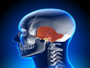
Anatomy of temporal bone
Technique
Step 1: Postauricular Incision and Exposure
A postauricular incision is made along the retroauricular groove. Expose mastoid cortex and posterior bony external auditory canal.
Step 2: Mastoidectomy and Removal of Temporal Bone Air Cells
Complete mastoidectomy with drilling of all air cells in the mastoid and petrous portion of the temporal bone.
Perform extended epitympanectomy for complete removal of middle ear air cells.
Step 3: Middle Ear and Eustachian Tube Management
Remove the tympanic membrane and ossicles. Close the eustachian tube with muscle or fascia to prevent reflux of nasopharyngeal contents.
Step 4: Facial Nerve and Inner Ear Preservation
Dissection near facial nerve with monitoring prevents injury; otic capsule preserved unless tumor invades.
Step 5: Cavity Obliteration
The mastoid cavity is filled with autologous tissue or synthetic materials to prevent dead space and CSF leakage.
Step 6: Closure
The temporalis muscle flap is used to cover the obliterated cavity. Multi-layered closure ensures watertight seal postauricular.
Subtotal petrosectomy (STP) is a surgical procedure that removes air cells from the temporal bone located near the skull base.
This procedure addresses chronic infections, CSF leaks, and ensures a stable surgical field for cochlear implants.
Temporal bone surgery evolved from radical mastoidectomy for infections treatment.
The development of the operating microscope and better anatomical understanding the emergence of STP for safe surgeries.
STP is crucial in lateral skull base surgery combined with procedures such as blind sac closure, cochlear implantation, and CSF otorrhea management.
Subtotal petrosectomy effectively manages complex otologic and skull base conditions, continually refined through advances in surgical techniques and imaging in otology and neurotology.
Chronic and Refractory Middle Ear Disease
Skull Base and Temporal Bone Tumors
Cerebrospinal Fluid (CSF) Leak and Encephalocele
Cochlear Implantation in Cases with Chronic Ear Disease
Palliative or Salvage Procedures
Post-Surgical Management and Revision Surgeries
Intact and functional hearing that must be preserved
Unstable or uncontrolled systemic conditions
Active intracranial infections
Severe coagulopathy
Extensive intracranial invasion by tumors
Patients with poor healing capacity
Patients unwilling to accept complete hearing loss
STP effectively treats chronic suppurative otitis media and recurrent issues.
Infection control exceeds 90% to reduce recurrent ear discharge and inflammation significantly.
STP prevents cholesteatoma reformation by eliminating air cells and sealing the external auditory canal.
Closure success rates for CSF otorrhea and encephaloceles exceed 85-90% with STP.
Patients receiving STP during cochlear implantation have success rates are similar with standard recipients.
Scalpel
Periosteal Elevator
Mastoid Retractors
Temporal Bone and Mastoidectomy Instruments
Middle Ear & Eustachian Tube Closure Instruments
Hemostasis & Cavity Obliteration Materials
Microsurgical & Neurovascular Instruments
Patient Preparation:
Administer general anesthesia to prevent nerve injury monitoring.
Ensures disease eradication that prevents recurrence.
Careful dissection around the facial nerve, dura, and inner ear structures.
Use fat, muscle, or synthetic materials to prevent dead space and CSF leakage.
Informed Consent:
Explain the procedure’s risks and potential complications clearly to the patient.
Patient Positioning:
Positioned patient in supine with head turned to the opposite side.
Anatomy of temporal bone
Step 1: Postauricular Incision and Exposure
A postauricular incision is made along the retroauricular groove. Expose mastoid cortex and posterior bony external auditory canal.
Step 2: Mastoidectomy and Removal of Temporal Bone Air Cells
Complete mastoidectomy with drilling of all air cells in the mastoid and petrous portion of the temporal bone.
Perform extended epitympanectomy for complete removal of middle ear air cells.
Step 3: Middle Ear and Eustachian Tube Management
Remove the tympanic membrane and ossicles. Close the eustachian tube with muscle or fascia to prevent reflux of nasopharyngeal contents.
Step 4: Facial Nerve and Inner Ear Preservation
Dissection near facial nerve with monitoring prevents injury; otic capsule preserved unless tumor invades.
Step 5: Cavity Obliteration
The mastoid cavity is filled with autologous tissue or synthetic materials to prevent dead space and CSF leakage.
Step 6: Closure
The temporalis muscle flap is used to cover the obliterated cavity. Multi-layered closure ensures watertight seal postauricular.
Both our subscription plans include Free CME/CPD AMA PRA Category 1 credits.
Digital Certificate PDF
On course completion, you will receive a full-sized presentation quality digital certificate.
medtigo Simulation
A dynamic medical simulation platform designed to train healthcare professionals and students to effectively run code situations through an immersive hands-on experience in a live, interactive 3D environment.
medtigo Points
medtigo points is our unique point redemption system created to award users for interacting on our site. These points can be redeemed for special discounts on the medtigo marketplace as well as towards the membership cost itself.
Community Forum post/reply = 5 points
*Redemption of points can occur only through the medtigo marketplace, courses, or simulation system. Money will not be credited to your bank account. 10 points = $1.
All Your Certificates in One Place
When you have your licenses, certificates and CMEs in one place, it's easier to track your career growth. You can easily share these with hospitals as well, using your medtigo app.