The sural nerve block is a regional anaesthetic technique which can be used as an adjunct to, or instead of, general anaesthesia for operations of the foot or ankle. This is relatively easy to perform because the sural nerve is located just below the skin around the ankle. The historical background of this technique can be traced back to 1965 where McCutcheon and colleagues pointed out a failure rate of 10%.
The sural nerve itself is not easily visualized when employing the ultrasound navigational technique, which is used in the new procedures of sural nerve blockage. Some of the factors that can increase the chances of success include perivascular techniques. Traditionally, an ankle block involves targeting four major nerves: The three main nerves that are usually blocked are the superficial peroneal, deep peroneal and the saphanous nerve as well as the tibial nerve together with the sural nerve block if necessary. However, it is recommended to include the sural nerve block in all cases not only to provide superior sensory anesthesia of the lateral foot and ankle region to minimize any inconvenience the patient might experience.
Indications
Indications for performing a sural nerve block include:
Examining or suture of wounds involving the lateral posterior calf or dorsolateral fifth finger.
A component of an ankle block required to treat a fractured or dislocated ankle.
Cleavage or evacuation of pus in abscess that forms in the posterolateral calf or in the laterodorsal fifth digit.
Surgical excision of a foreign object from the postero-lateral calf or dorsolateral fifth toe.
Contraindications
Contraindications for a sural nerve block include:
Known hypersensitivity to anesthetic drugs or any of its components (e.g. ester or amide type).
Injection through infected tissue.
Severe bleeding disorders or coagulopathy such as haemophilia, thrombocytopenia, leukaemia, or liver disease.
Existing neurological damage.
Failure to get patient cooperation (some patients especially pediatric or elderly ones may need to be anesthetized).
Outcomes
Equipment
Antiseptic Solution: povidone iodine or Chlorhexidine gluconate
Ultrasound Equipment: Ultrasound probe with high frequency with sterile gel
Local Anaesthetic: Usually, a 1% lidocaine solution for superficial block of the skin.
Regional Anaesthetic Solution: 0.5% ropivacaine or bupivacaine for longer acting analgesia and 2% of lidocaine or 1.5% of mepivacaine as fast acting agents.
Syringe: A 10 to 20 mL of syringe along with a piece of extension tubing.
Needle: Block needle of 2 inch in length with 22 gauge.
Patient preparation
First seek consent from the patient and ensure that the patient understands all the procedures to be taken. After a pause before the procedure, the patient is placed to expose and provide access to the lateral side of the leg. Propofol and midazolam can be used for sedation while fentanyl may be used for analgesia. Aseptic procedure is observed, including the use of 2% of chlorhexidine gluconate or povidone-iodine solution at the site of the injection. The probe used during the ultrasound is then disinfected using a sterile probe cover and then wiped with sterile gel.
Patient position
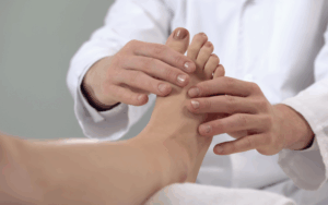
Place the patient in a prone position whereby the ankle part should be at a comfortable position through being supported and raised with a pillow or a rolled sheet.
Another position of the patient is seated or lying down on the back with the involved limb in a position of internal rotation and the foot elevated on a pillow or a rolled sheet.
To ensure the success of the procedure, it is vital to prepare the equipment correctly and position the patient in the right manner.
Patient positioning with sural nerve block
Technique
Ultrasound-Guided Approach:
Step 1-Identify the base of the lateral malleolus behind and extending down as far as the Achilles tendon.
Step 2– Identification of the site where the mark must be made; it is the site that is positioned laterally to the Achilles tendon and posterior mediolaterally located to the lateral side of the malleolus.
Step 3-Laterally position the linear high frequency between 10 to 18 mega Hertz ultrasound transducer at the mark.
The first and the foremost step within this strategy includes guiding the ultrasound probe to locate the small saphenous vein.
Step 5-Identification of injection technique:
Out of the Plane Approach: Administer about 5 ml of the local anaesthetic in a peripheral fashion to the area surrounding the lesser saphenous vein.
In-Plane Approach: Proceeding with needle from the tip to the base along the line pointing to the sural nerve in the line parallel to the length of the limb.
Step 6-Incase the leg or uplifting the tourniquet of the calf to a higher level to make the veins larger to find sural nerve easily.
Landmark-Based Approach:
Step 1-Determine the position of the two points, the knot between the two lobes of the Achilles tendon and where one lobe of this tendon touches the backside of the lateral side of the malleolus.
Step 2-The puncture should be made proximal to the distal tip of the needle, approximately at a level superior to the tip of the lateral malleolus joint line resting on the Achilles tendon laterally.
Step 3-In the case of paraesthesia, do not advance the needle anymore, conduct negative aspiration test and inject 3 to 5 mL of local anesthetic.
Step 4-If paresthesia is not encountered then the needle is made to move further until it touches the lateral malleolus bone and then the local anesthetic is administered while withdrawing the needle.
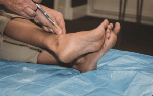
Sural Nerve block-Administration of local anaesthetics
Approach considerations
Perform neurovascular studies and evaluate sural nerve sensation.
Identify the area lateral to the tendon Achilles approximately 1 inch proximal to the lateral malleolus.
Clean the area with disinfectant and get rid of all the contaminated appliances and apparatus.
Prepare lidocaine using an 18-gauge needle and then change it to a 25-gauge one.
Puncture the skin to make a wheal and push the needle forward in the direction of the lateral malleolus and inject the lidocaine 5-7 ml.
Evaluate for anesthesia and observe for alterations in foot color or temperature.
Laboratory tests
Complete blood count
Renal function test
Coagulation profile
Complications
Side effects are extremely unlikely but include pain at the injection site, bruising, infection, and allergic reactions. Local anaesthetic toxicity occurs when intravascular injection is made. Furthermore, moderate to severe pain, nerve injury, and tissue damage occur when the needle tip is placed subfascicularly, perineurally, or intraneurally.
The sural nerve block is a regional anaesthetic technique which can be used as an adjunct to, or instead of, general anaesthesia for operations of the foot or ankle. This is relatively easy to perform because the sural nerve is located just below the skin around the ankle. The historical background of this technique can be traced back to 1965 where McCutcheon and colleagues pointed out a failure rate of 10%.
The sural nerve itself is not easily visualized when employing the ultrasound navigational technique, which is used in the new procedures of sural nerve blockage. Some of the factors that can increase the chances of success include perivascular techniques. Traditionally, an ankle block involves targeting four major nerves: The three main nerves that are usually blocked are the superficial peroneal, deep peroneal and the saphanous nerve as well as the tibial nerve together with the sural nerve block if necessary. However, it is recommended to include the sural nerve block in all cases not only to provide superior sensory anesthesia of the lateral foot and ankle region to minimize any inconvenience the patient might experience.
Indications for performing a sural nerve block include:
Examining or suture of wounds involving the lateral posterior calf or dorsolateral fifth finger.
A component of an ankle block required to treat a fractured or dislocated ankle.
Cleavage or evacuation of pus in abscess that forms in the posterolateral calf or in the laterodorsal fifth digit.
Surgical excision of a foreign object from the postero-lateral calf or dorsolateral fifth toe.
Contraindications for a sural nerve block include:
Known hypersensitivity to anesthetic drugs or any of its components (e.g. ester or amide type).
Injection through infected tissue.
Severe bleeding disorders or coagulopathy such as haemophilia, thrombocytopenia, leukaemia, or liver disease.
Existing neurological damage.
Failure to get patient cooperation (some patients especially pediatric or elderly ones may need to be anesthetized).
Antiseptic Solution: povidone iodine or Chlorhexidine gluconate
Ultrasound Equipment: Ultrasound probe with high frequency with sterile gel
Local Anaesthetic: Usually, a 1% lidocaine solution for superficial block of the skin.
Regional Anaesthetic Solution: 0.5% ropivacaine or bupivacaine for longer acting analgesia and 2% of lidocaine or 1.5% of mepivacaine as fast acting agents.
Syringe: A 10 to 20 mL of syringe along with a piece of extension tubing.
Needle: Block needle of 2 inch in length with 22 gauge.
First seek consent from the patient and ensure that the patient understands all the procedures to be taken. After a pause before the procedure, the patient is placed to expose and provide access to the lateral side of the leg. Propofol and midazolam can be used for sedation while fentanyl may be used for analgesia. Aseptic procedure is observed, including the use of 2% of chlorhexidine gluconate or povidone-iodine solution at the site of the injection. The probe used during the ultrasound is then disinfected using a sterile probe cover and then wiped with sterile gel.
Place the patient in a prone position whereby the ankle part should be at a comfortable position through being supported and raised with a pillow or a rolled sheet.
Another position of the patient is seated or lying down on the back with the involved limb in a position of internal rotation and the foot elevated on a pillow or a rolled sheet.
To ensure the success of the procedure, it is vital to prepare the equipment correctly and position the patient in the right manner.
Patient positioning with sural nerve block
Ultrasound-Guided Approach:
Step 1-Identify the base of the lateral malleolus behind and extending down as far as the Achilles tendon.
Step 2– Identification of the site where the mark must be made; it is the site that is positioned laterally to the Achilles tendon and posterior mediolaterally located to the lateral side of the malleolus.
Step 3-Laterally position the linear high frequency between 10 to 18 mega Hertz ultrasound transducer at the mark.
The first and the foremost step within this strategy includes guiding the ultrasound probe to locate the small saphenous vein.
Step 5-Identification of injection technique:
Out of the Plane Approach: Administer about 5 ml of the local anaesthetic in a peripheral fashion to the area surrounding the lesser saphenous vein.
In-Plane Approach: Proceeding with needle from the tip to the base along the line pointing to the sural nerve in the line parallel to the length of the limb.
Step 6-Incase the leg or uplifting the tourniquet of the calf to a higher level to make the veins larger to find sural nerve easily.
Landmark-Based Approach:
Step 1-Determine the position of the two points, the knot between the two lobes of the Achilles tendon and where one lobe of this tendon touches the backside of the lateral side of the malleolus.
Step 2-The puncture should be made proximal to the distal tip of the needle, approximately at a level superior to the tip of the lateral malleolus joint line resting on the Achilles tendon laterally.
Step 3-In the case of paraesthesia, do not advance the needle anymore, conduct negative aspiration test and inject 3 to 5 mL of local anesthetic.
Step 4-If paresthesia is not encountered then the needle is made to move further until it touches the lateral malleolus bone and then the local anesthetic is administered while withdrawing the needle.
Sural Nerve block-Administration of local anaesthetics
Perform neurovascular studies and evaluate sural nerve sensation.
Identify the area lateral to the tendon Achilles approximately 1 inch proximal to the lateral malleolus.
Clean the area with disinfectant and get rid of all the contaminated appliances and apparatus.
Prepare lidocaine using an 18-gauge needle and then change it to a 25-gauge one.
Puncture the skin to make a wheal and push the needle forward in the direction of the lateral malleolus and inject the lidocaine 5-7 ml.
Evaluate for anesthesia and observe for alterations in foot color or temperature.
Complete blood count
Renal function test
Coagulation profile
Side effects are extremely unlikely but include pain at the injection site, bruising, infection, and allergic reactions. Local anaesthetic toxicity occurs when intravascular injection is made. Furthermore, moderate to severe pain, nerve injury, and tissue damage occur when the needle tip is placed subfascicularly, perineurally, or intraneurally.
Both our subscription plans include Free CME/CPD AMA PRA Category 1 credits.
Digital Certificate PDF
On course completion, you will receive a full-sized presentation quality digital certificate.
medtigo Simulation
A dynamic medical simulation platform designed to train healthcare professionals and students to effectively run code situations through an immersive hands-on experience in a live, interactive 3D environment.
medtigo Points
medtigo points is our unique point redemption system created to award users for interacting on our site. These points can be redeemed for special discounts on the medtigo marketplace as well as towards the membership cost itself.
Community Forum post/reply = 5 points
*Redemption of points can occur only through the medtigo marketplace, courses, or simulation system. Money will not be credited to your bank account. 10 points = $1.
All Your Certificates in One Place
When you have your licenses, certificates and CMEs in one place, it's easier to track your career growth. You can easily share these with hospitals as well, using your medtigo app.