Background
Anal fissures are small tears in the lining of the anal canal, often caused by trauma during bowel movements or conditions leading to increased anal sphincter tone. They can cause significant pain, bleeding, and discomfort. While many anal fissures resolve with conservative treatments like dietary changes, topical medications, and stool softeners, chronic or refractory cases may require surgical intervention.
Surgical management is aimed at relieving sphincter spasm, promoting healing, and preventing recurrence while minimizing complications such as incontinence. The two primary surgical options are lateral internal sphincterotomy (LIS) and newer techniques such as botulinum toxin injection or fissurectomy in specific cases. LIS is widely regarded as the gold standard for chronic anal fissures due to its high success and low recurrence rates.
Indications
Chronic Anal Fissure:
Persistence of symptoms (pain, bleeding, discomfort) for more than 6–8 weeks despite conservative treatments, such as dietary modifications, stool softeners, sitz baths, and topical medications (e.g., nitro-glycerine or calcium channel blockers).
Recurrent Fissures:
Patients experiencing multiple episodes of anal fissures over time that do not resolve with non-surgical treatments.
Severe Pain:
Intolerable pain affecting daily activities and quality of life, unrelieved by medications and other conservative measures.
Complicated Anal Fissure:
Fissures associated with:
Significant scarring or fibrosis.
Development of skin tags or sentinel piles.
Anal stenosis.
Fistula formation or infection.
Underlying Disorders:
When the fissure is secondary to conditions like Crohn’s disease or malignancy, surgery might be part of a broader treatment plan, though the underlying condition must also be addressed.
Failure of Conservative Therapy:
No improvement after 6–12 weeks of appropriate non-surgical management.
Contraindications
Active Infections:
Perianal or rectal infections (e.g., abscess or fistula): Surgery should be delayed until the infection is treated.
Underlying Conditions:
Inflammatory Bowel Disease (IBD): Conditions like Crohn’s disease or ulcerative colitis may require alternative treatments due to impaired healing and increased risk of complications.
Malignancy: Suspected or confirmed anal or rectal cancer should be addressed with appropriate oncologic evaluation and treatment.
Coagulopathies or Anticoagulation:
Patients with uncontrolled bleeding disorders or those on anticoagulant therapy that cannot be safely discontinued may have an increased risk of postoperative bleeding.
Poor Healing Conditions:
Diabetes mellitus with poor glycemic control: Increased risk of infection and delayed wound healing.
Malnutrition or other systemic conditions impairing wound healing.
Functional or Structural Concerns:
Anal incontinence: Lateral internal sphincterotomy may worsen pre-existing incontinence.
Pelvic floor dysfunction: May complicate outcomes and recovery.
Outcomes
Equipment
Surgical Instruments:
Scalpels (#15 blade or similar)
Fine scissors (e.g., Metzenbaum scissors)
Small retractors (e.g., anal retractors)
Needle holders
Fine forceps (toothed and non-toothed)
Anoscopy Equipment:

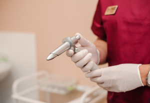
Anoscope
Anoscope (to visualize the anal canal)
Electrocautery or Diathermy
Sutures and Needles
Absorbable sutures
Hemostatic Materials
Gauze or sponges for hemostasis.
Hemostatic agents (e.g., Surgicel or Gelfoam).
Local Anesthetic and Syringes
Needles (23G or finer for precise application).
Patient preparation
Pre-Surgery Preparation
Medical Evaluation:
The surgeon will conduct a detailed medical history review and physical examination.
Any comorbid conditions (e.g., diabetes, hypertension) will need to be stabilized before surgery.
Medications:
Disclose all medications, supplements, and herbal remedies to the surgeon.
You may need to stop blood-thinning medications (e.g., aspirin, warfarin, clopidogrel) several days prior, under the guidance of your doctor.
Bowel Preparation:
Some surgeons may recommend a gentle enema or a bowel-cleansing solution before surgery to clear the rectum.
Avoid heavy or fatty meals the night before surgery.
Dietary Restrictions:
Patients are typically advised to fast (no food or drink) for 6–8 hours before the surgery, depending on anesthesia guidelines.
Patient position
Prone Jackknife Position:
The patient lies on their abdomen, with the hips elevated and the legs lowered.
This position allows direct access to the anal area and is preferred for certain surgeons due to ease of exposure and reduced pressure on the perineum.
Left Lateral (Sims’) Position:
The patient lies on their left side, with the right hip and knee flexed (left leg extended or slightly flexed).
This position is common for minor anorectal procedures performed under local or regional anesthesia.
Lateral Internal Sphincterotomy
Step 1-Positioning:
The patient is placed in the lithotomy position (on the back with legs elevated and spread apart) to allow good access to the anal canal.
The area around the anus is cleaned and sterilized to prevent infection.
Step 2-Anoscopy and Identification of the Fissure:
Insert an anal speculum (anoscope) into the anal canal to visualize the fissure, its location, and any associated abnormalities (e.g., hypertrophied anal papillae, sentinel tag).
The internal anal sphincter is located just below the dentate line, and the fissure is usually at the posterior or anterior midline.
Step 3- Injection of Local Anesthesia (Optional):
In some cases, local anesthesia (such as 1-2% lidocaine with epinephrine) is injected around the sphincter to ensure proper analgesia. This can be done in addition to general anesthesia if needed.
Step 4-Incision of the Internal Sphincter:
A lateral incision is made in the internal anal sphincter muscle. The incision is usually about 1–2 cm in length and is made at the 6 o’clock position (posterior midline) for posterior fissures or at the 3 o’clock or 9 o’clock position for anterior fissures.
The incision is deep enough to divide the muscle but not too deep to damage the external sphincter or cause incontinence.
Step 5-Muscle Division:
The internal anal sphincter is carefully divided in a lateral direction using scissors or a scalpel.
The goal is to reduce the pressure within the anal canal, relieving the spasm that contributes to the fissure.
Step 6-Hemostasis:
Ensure there is no significant bleeding after the incision. If necessary, use electrocautery or sutures to control bleeding.
Step 7-Wound Closure:
Typically, the incision in the sphincter does not require suturing. However, if there is significant bleeding or other concerns, a simple absorbable suture may be used to control bleeding.
Step 8-Postoperative Care:
Postoperative Pain Management: Prescribe pain relievers (e.g., acetaminophen or ibuprofen) and possibly a topical anesthetic.
Sitz Baths: Encourage warm sitz baths to help relax the anal muscles and promote healing.
Laxatives: Recommend stool softeners to prevent straining during bowel movements.
Follow-up: Schedule a follow-up appointment to monitor for complications such as infection, incontinence, or persistent fissures.
Fissurectomy
Step 1-Positioning:
Place the patient in a lithotomy or prone jackknife position (depending on surgeon preference).
Expose the anal region for a clear view of the fissure.
Step 2-Anoscopy:
Use an anoscope to visualize the anal canal and locate the fissure accurately.
Step 3-Fissure Identification:
Identify the fissure, which usually appears as a crack or tear in the anoderm.
Check for signs of infection, scarring, or abscess formation.
Step 4-Incision and Removal:
Carefully excise the fissure and any surrounding scar tissue, ensuring that the entire fissure is removed without damaging healthy tissue.
The surgeon may also remove any excess fibrous tissue or redundant skin that has formed due to chronic fissures.
Step 5-Hemostasis:
Ensure there is no active bleeding at the surgical site. If necessary, cauterize or use sutures to control bleeding.
Step 6-Sphincterotomy (optional):
In some cases, a partial sphincterotomy (cutting part of the internal anal sphincter) is performed to relieve excessive sphincter tension, which can promote fissure healing. This step is particularly common in chronic or recurrent fissures.
Step 7-Wound Closure:
Leave the wound open or use absorbable sutures if necessary, depending on the surgical approach and type of fissure being treated.
Step 8-Postoperative Care:
Pain Management:
Administer pain relief medication, typically nonsteroidal anti-inflammatory drugs (NSAIDs) or opioids if necessary.
Prescribe stool softeners to minimize straining during bowel movements.
Follow-up:
Schedule a follow-up appointment to assess the healing process and check for complications, such as infection or recurrence of fissures.
Complications
Pain:
Post-surgical pain is common and usually manageable with medication.
Severe or prolonged pain may indicate infection or other issues.
Infection
Infection at the surgical site is possible. Symptoms may include redness, swelling, warmth, or discharge.
Prompt medical attention is necessary if infection is suspected.
Bleeding
Minor bleeding can occur post-surgery but usually subsides within a few days.
Persistent or heavy bleeding may require medical evaluation.
Incontinence
Gas incontinence: Difficulty controlling gas is the most common.
Fecal incontinence: Rare but may occur, especially if too much of the sphincter muscle is cut.
Abscess or Fistula Formation
Rarely, an infection may develop into an abscess, or an abnormal connection (fistula) may form between the anal canal and surrounding tissues.
Delayed Healing
Healing time varies, but some patients may experience delayed healing of the fissure or surgical wound.
Recurrence of the Fissure
The fissure may reoccur in a small percentage of cases, requiring further treatment.
Scar Tissue Formation
Scar tissue at the surgical site may cause discomfort or narrowing of the anal canal in rare cases.
References
References

Anal fissures are small tears in the lining of the anal canal, often caused by trauma during bowel movements or conditions leading to increased anal sphincter tone. They can cause significant pain, bleeding, and discomfort. While many anal fissures resolve with conservative treatments like dietary changes, topical medications, and stool softeners, chronic or refractory cases may require surgical intervention.
Surgical management is aimed at relieving sphincter spasm, promoting healing, and preventing recurrence while minimizing complications such as incontinence. The two primary surgical options are lateral internal sphincterotomy (LIS) and newer techniques such as botulinum toxin injection or fissurectomy in specific cases. LIS is widely regarded as the gold standard for chronic anal fissures due to its high success and low recurrence rates.
Chronic Anal Fissure:
Persistence of symptoms (pain, bleeding, discomfort) for more than 6–8 weeks despite conservative treatments, such as dietary modifications, stool softeners, sitz baths, and topical medications (e.g., nitro-glycerine or calcium channel blockers).
Recurrent Fissures:
Patients experiencing multiple episodes of anal fissures over time that do not resolve with non-surgical treatments.
Severe Pain:
Intolerable pain affecting daily activities and quality of life, unrelieved by medications and other conservative measures.
Complicated Anal Fissure:
Fissures associated with:
Significant scarring or fibrosis.
Development of skin tags or sentinel piles.
Anal stenosis.
Fistula formation or infection.
Underlying Disorders:
When the fissure is secondary to conditions like Crohn’s disease or malignancy, surgery might be part of a broader treatment plan, though the underlying condition must also be addressed.
Failure of Conservative Therapy:
No improvement after 6–12 weeks of appropriate non-surgical management.
Active Infections:
Perianal or rectal infections (e.g., abscess or fistula): Surgery should be delayed until the infection is treated.
Underlying Conditions:
Inflammatory Bowel Disease (IBD): Conditions like Crohn’s disease or ulcerative colitis may require alternative treatments due to impaired healing and increased risk of complications.
Malignancy: Suspected or confirmed anal or rectal cancer should be addressed with appropriate oncologic evaluation and treatment.
Coagulopathies or Anticoagulation:
Patients with uncontrolled bleeding disorders or those on anticoagulant therapy that cannot be safely discontinued may have an increased risk of postoperative bleeding.
Poor Healing Conditions:
Diabetes mellitus with poor glycemic control: Increased risk of infection and delayed wound healing.
Malnutrition or other systemic conditions impairing wound healing.
Functional or Structural Concerns:
Anal incontinence: Lateral internal sphincterotomy may worsen pre-existing incontinence.
Pelvic floor dysfunction: May complicate outcomes and recovery.
Surgical Instruments:
Scalpels (#15 blade or similar)
Fine scissors (e.g., Metzenbaum scissors)
Small retractors (e.g., anal retractors)
Needle holders
Fine forceps (toothed and non-toothed)
Anoscopy Equipment:

Anoscope
Anoscope (to visualize the anal canal)
Electrocautery or Diathermy
Sutures and Needles
Absorbable sutures
Hemostatic Materials
Gauze or sponges for hemostasis.
Hemostatic agents (e.g., Surgicel or Gelfoam).
Local Anesthetic and Syringes
Needles (23G or finer for precise application).
Patient preparation
Pre-Surgery Preparation
Medical Evaluation:
The surgeon will conduct a detailed medical history review and physical examination.
Any comorbid conditions (e.g., diabetes, hypertension) will need to be stabilized before surgery.
Medications:
Disclose all medications, supplements, and herbal remedies to the surgeon.
You may need to stop blood-thinning medications (e.g., aspirin, warfarin, clopidogrel) several days prior, under the guidance of your doctor.
Bowel Preparation:
Some surgeons may recommend a gentle enema or a bowel-cleansing solution before surgery to clear the rectum.
Avoid heavy or fatty meals the night before surgery.
Dietary Restrictions:
Patients are typically advised to fast (no food or drink) for 6–8 hours before the surgery, depending on anesthesia guidelines.
Patient position
Prone Jackknife Position:
The patient lies on their abdomen, with the hips elevated and the legs lowered.
This position allows direct access to the anal area and is preferred for certain surgeons due to ease of exposure and reduced pressure on the perineum.
Left Lateral (Sims’) Position:
The patient lies on their left side, with the right hip and knee flexed (left leg extended or slightly flexed).
This position is common for minor anorectal procedures performed under local or regional anesthesia.
Step 1-Positioning:
The patient is placed in the lithotomy position (on the back with legs elevated and spread apart) to allow good access to the anal canal.
The area around the anus is cleaned and sterilized to prevent infection.
Step 2-Anoscopy and Identification of the Fissure:
Insert an anal speculum (anoscope) into the anal canal to visualize the fissure, its location, and any associated abnormalities (e.g., hypertrophied anal papillae, sentinel tag).
The internal anal sphincter is located just below the dentate line, and the fissure is usually at the posterior or anterior midline.
Step 3- Injection of Local Anesthesia (Optional):
In some cases, local anesthesia (such as 1-2% lidocaine with epinephrine) is injected around the sphincter to ensure proper analgesia. This can be done in addition to general anesthesia if needed.
Step 4-Incision of the Internal Sphincter:
A lateral incision is made in the internal anal sphincter muscle. The incision is usually about 1–2 cm in length and is made at the 6 o’clock position (posterior midline) for posterior fissures or at the 3 o’clock or 9 o’clock position for anterior fissures.
The incision is deep enough to divide the muscle but not too deep to damage the external sphincter or cause incontinence.
Step 5-Muscle Division:
The internal anal sphincter is carefully divided in a lateral direction using scissors or a scalpel.
The goal is to reduce the pressure within the anal canal, relieving the spasm that contributes to the fissure.
Step 6-Hemostasis:
Ensure there is no significant bleeding after the incision. If necessary, use electrocautery or sutures to control bleeding.
Step 7-Wound Closure:
Typically, the incision in the sphincter does not require suturing. However, if there is significant bleeding or other concerns, a simple absorbable suture may be used to control bleeding.
Step 8-Postoperative Care:
Postoperative Pain Management: Prescribe pain relievers (e.g., acetaminophen or ibuprofen) and possibly a topical anesthetic.
Sitz Baths: Encourage warm sitz baths to help relax the anal muscles and promote healing.
Laxatives: Recommend stool softeners to prevent straining during bowel movements.
Follow-up: Schedule a follow-up appointment to monitor for complications such as infection, incontinence, or persistent fissures.
Step 1-Positioning:
Place the patient in a lithotomy or prone jackknife position (depending on surgeon preference).
Expose the anal region for a clear view of the fissure.
Step 2-Anoscopy:
Use an anoscope to visualize the anal canal and locate the fissure accurately.
Step 3-Fissure Identification:
Identify the fissure, which usually appears as a crack or tear in the anoderm.
Check for signs of infection, scarring, or abscess formation.
Step 4-Incision and Removal:
Carefully excise the fissure and any surrounding scar tissue, ensuring that the entire fissure is removed without damaging healthy tissue.
The surgeon may also remove any excess fibrous tissue or redundant skin that has formed due to chronic fissures.
Step 5-Hemostasis:
Ensure there is no active bleeding at the surgical site. If necessary, cauterize or use sutures to control bleeding.
Step 6-Sphincterotomy (optional):
In some cases, a partial sphincterotomy (cutting part of the internal anal sphincter) is performed to relieve excessive sphincter tension, which can promote fissure healing. This step is particularly common in chronic or recurrent fissures.
Step 7-Wound Closure:
Leave the wound open or use absorbable sutures if necessary, depending on the surgical approach and type of fissure being treated.
Step 8-Postoperative Care:
Pain Management:
Administer pain relief medication, typically nonsteroidal anti-inflammatory drugs (NSAIDs) or opioids if necessary.
Prescribe stool softeners to minimize straining during bowel movements.
Follow-up:
Schedule a follow-up appointment to assess the healing process and check for complications, such as infection or recurrence of fissures.
Complications
Pain:
Post-surgical pain is common and usually manageable with medication.
Severe or prolonged pain may indicate infection or other issues.
Infection
Infection at the surgical site is possible. Symptoms may include redness, swelling, warmth, or discharge.
Prompt medical attention is necessary if infection is suspected.
Bleeding
Minor bleeding can occur post-surgery but usually subsides within a few days.
Persistent or heavy bleeding may require medical evaluation.
Incontinence
Gas incontinence: Difficulty controlling gas is the most common.
Fecal incontinence: Rare but may occur, especially if too much of the sphincter muscle is cut.
Abscess or Fistula Formation
Rarely, an infection may develop into an abscess, or an abnormal connection (fistula) may form between the anal canal and surrounding tissues.
Delayed Healing
Healing time varies, but some patients may experience delayed healing of the fissure or surgical wound.
Recurrence of the Fissure
The fissure may reoccur in a small percentage of cases, requiring further treatment.
Scar Tissue Formation
Scar tissue at the surgical site may cause discomfort or narrowing of the anal canal in rare cases.

Both our subscription plans include Free CME/CPD AMA PRA Category 1 credits.

On course completion, you will receive a full-sized presentation quality digital certificate.
A dynamic medical simulation platform designed to train healthcare professionals and students to effectively run code situations through an immersive hands-on experience in a live, interactive 3D environment.

When you have your licenses, certificates and CMEs in one place, it's easier to track your career growth. You can easily share these with hospitals as well, using your medtigo app.


