Tracheal Intubation

Updated : September 4, 2024

Mail Whatsapp PDF Image

Background

Tracheal intubation is a medical procedure in which a flexible tube is inserted into the trachea.

Tracheal tubes vary in size and design for different patients with cuffs help aspiration prevention and ensure good ventilation.

Direct laryngoscopy is used as a traditional method in which a laryngoscope sees vocal cords for intubation.

Procedure conducted using direct or video laryngoscopy guidance and selection of technique are based on clinical scenario and anatomy of patient.

Indications

Tracheal intubation indicated for upper airway blockage including angioedema and epiglottitis.

Intubation used in respiratory failure cases, including acute respiratory distress syndrome (ARDS) treatment for patient.

Patients with less consciousness level may need this procedure to protect airway.

Tracheal intubation indicated during cardiopulmonary resuscitation to give open airway.

Contraindications

Tracheal intubation is not performed in cardiac arrest with no chance of recovery.

It should be contraindicated in patients with skull fractures because it causes cerebrospinal fluid leakage.

In severe hypoxemia or hemodynamic instability cases, the risks associated with intubation may beat the potential benefits.

Patients with intact protective airway reflexes may not need intubation unless necessary.

Outcomes

It controls oxygen supply and ventilation, which improves oxygen level in patients.

Intubation prevents aspiration, thus decreases risk of pneumonia, and minimizes complications.

Monitor intubated patients for respiratory parameters such as oxygen saturation and CO2.

Intubation indicates resolved condition or improved respiratory function, which is required in ongoing support for smooth recovery.

Equipment

  • Laryngoscope
  • Endotracheal Tubes
  • Stylet
  • Suction Apparatus
  • End-tidal Carbon Dioxide Monitoring
  • Oropharyngeal and Nasopharyngeal Airway Devices

Patient Preparation

Informed Consent:

The procedure should be thoroughly discussed with the patient, to ensure informed consent.

 Patient Positioning:

Place patient in optimal airway alignment, usually with head in sniffing position for vocal cord visualization.

Technique

Step 1: Anesthesia

Inject sedatives as anesthesia for relaxing muscles. Before this ensure proper supply of oxygen for patient during intubation.

Step 2: Vocal Cord Visualization

The surgeon inserted laryngoscope in mouth, then slowly displaced to see the epiglottis.

Lift epiglottis to expose vocal cords for visualization.

Step 3: Tube insertion

Insert endotracheal tube through mouth along tongue curve then guide towards glottis.

Use correct air volume of cuff endotracheal tube to prevent aspiration.

                             Fig. Anterior neck view

Step 4: Confirmation

Confirm proper endotracheal tube placement with help of chest X-ray and listening stable breath sounds.

Complications

Intubation has risks of introducing pathogens, which increase pneumonia in respiratory patients.

Misuse of laryngoscope blade and endotracheal tube insertion may damage larynx and trachea.

Misplacement of endotracheal tube can lead to insufficient breathing support.

Incorrect tube placement or cuff inflation can lead to gastric aspiration.

Content loading

Tracheal Intubation

Updated : September 4, 2024

Mail Whatsapp PDF Image


Tracheal intubation is a medical procedure in which a flexible tube is inserted into the trachea.

Tracheal tubes vary in size and design for different patients with cuffs help aspiration prevention and ensure good ventilation.

Direct laryngoscopy is used as a traditional method in which a laryngoscope sees vocal cords for intubation.

Procedure conducted using direct or video laryngoscopy guidance and selection of technique are based on clinical scenario and anatomy of patient.

Tracheal intubation indicated for upper airway blockage including angioedema and epiglottitis.

Intubation used in respiratory failure cases, including acute respiratory distress syndrome (ARDS) treatment for patient.

Patients with less consciousness level may need this procedure to protect airway.

Tracheal intubation indicated during cardiopulmonary resuscitation to give open airway.

Tracheal intubation is not performed in cardiac arrest with no chance of recovery.

It should be contraindicated in patients with skull fractures because it causes cerebrospinal fluid leakage.

In severe hypoxemia or hemodynamic instability cases, the risks associated with intubation may beat the potential benefits.

Patients with intact protective airway reflexes may not need intubation unless necessary.

It controls oxygen supply and ventilation, which improves oxygen level in patients.

Intubation prevents aspiration, thus decreases risk of pneumonia, and minimizes complications.

Monitor intubated patients for respiratory parameters such as oxygen saturation and CO2.

Intubation indicates resolved condition or improved respiratory function, which is required in ongoing support for smooth recovery.

  • Laryngoscope
  • Endotracheal Tubes
  • Stylet
  • Suction Apparatus
  • End-tidal Carbon Dioxide Monitoring
  • Oropharyngeal and Nasopharyngeal Airway Devices

Informed Consent:

The procedure should be thoroughly discussed with the patient, to ensure informed consent.

 Patient Positioning:

Place patient in optimal airway alignment, usually with head in sniffing position for vocal cord visualization.

Step 1: Anesthesia

Inject sedatives as anesthesia for relaxing muscles. Before this ensure proper supply of oxygen for patient during intubation.

Step 2: Vocal Cord Visualization

The surgeon inserted laryngoscope in mouth, then slowly displaced to see the epiglottis.

Lift epiglottis to expose vocal cords for visualization.

Step 3: Tube insertion

Insert endotracheal tube through mouth along tongue curve then guide towards glottis.

Use correct air volume of cuff endotracheal tube to prevent aspiration.

                             Fig. Anterior neck view

Step 4: Confirmation

Confirm proper endotracheal tube placement with help of chest X-ray and listening stable breath sounds.

Intubation has risks of introducing pathogens, which increase pneumonia in respiratory patients.

Misuse of laryngoscope blade and endotracheal tube insertion may damage larynx and trachea.

Misplacement of endotracheal tube can lead to insufficient breathing support.

Incorrect tube placement or cuff inflation can lead to gastric aspiration.

Free CME credits

Both our subscription plans include Free CME/CPD AMA PRA Category 1 credits.

Digital Certificate PDF

On course completion, you will receive a full-sized presentation quality digital certificate.
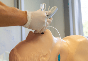
medtigo Simulation

A dynamic medical simulation platform designed to train healthcare professionals and students to effectively run code situations through an immersive hands-on experience in a live, interactive 3D environment.

medtigo Points

medtigo points is our unique point redemption system created to award users for interacting on our site. These points can be redeemed for special discounts on the medtigo marketplace as well as towards the membership cost itself.
 
  • Registration with medtigo = 10 points
  • 1 visit to medtigo’s website = 1 point
  • Interacting with medtigo posts (through comments/clinical cases etc.) = 5 points
  • Attempting a game = 1 point
  • Community Forum post/reply = 5 points

    *Redemption of points can occur only through the medtigo marketplace, courses, or simulation system. Money will not be credited to your bank account. 10 points = $1.

All Your Certificates in One Place

When you have your licenses, certificates and CMEs in one place, it's easier to track your career growth. You can easily share these with hospitals as well, using your medtigo app.

Our Certificate Courses