Background
Transthoracic echocardiography (TTE) is non-invasive method to evaluate heart structure and function.
It is commonly used diagnostic tools in cardiology that provide real-time images of the heart using ultrasound waves.
TTE assesses heart function and identifies cardiac symptom causes. The test reveals heart chambers, valves, and surrounding blood vessels.
Echocardiography uses ultrasound waves for body imaging. Computer technology visualizes heart images using echoes from heartbeats.
Doppler ultrasound in echocardiograms assesses blood flow in heart chambers and valves.
It shows portability, safety, availability, and non-invasive assessment of heart morphology and physiology.
Many patients receive prior TTE before advanced imaging with CT or MRI for cardiac procedures.
A phased-array transducer is chosen for TTE, using multiple elements fired sequentially to generate a sector image with a wide field of view.
Indications
Evaluation of Cardiac Symptoms
Assessment of Left and Right Ventricular Function
Valvular Heart Disease
Infective Endocarditis
Pericardial Disease
Congenital Heart Disease
Aortic Disease
Embolic Events
Contraindications
Poor Acoustic Windows
Inability to Maintain Proper Positioning
Presence of Subcutaneous Emphysema
Esophageal or Gastric Surgery
Open Chest Wounds
Outcomes
Evaluates ventricular relaxation issues in HFpEF diagnosis. TTE confirms or rules out suspected cardiac conditions with details.
Evaluates pulmonary hypertension and right heart failure conditions.
It identifies thrombi in atrial fibrillation and myocardial infarction. Severity assessment guides surgical intervention decisions.
Obesity and lung disease decrease diagnostic accuracy and image quality.
Outcomes vary by clinical indication, imaging quality, and management decisions post-findings.
Equipment required
Echocardiography Machine
Transducer
Ultrasound Gel
Electrocardiogram Leads
Software & Image Processing Tools
Patient Preparation:
No fasting is required during TTE method. Patients should continue with their daily medications.
Patients need to remove upper cloths and wear a hospital gown for chest access.
Apply ultrasound gel on the probe to improve sound wave transmission.
Informed Consent:
Explain the procedure’s risks and potential complications clearly to the patient.
Patient Positioning:
Use left lateral decubitus for heart proximity and in supine positioned if left lateral is intolerable.

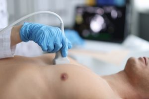
Figure. Transthoracic echocardiography
Step 1: Probe Selection and Placement
Select transducer type as phased-array transducer for deep penetration and cardiac imaging.
Standard probe positions are:
Parasternal window for Left side of the sternum
Apical window for near the apex of the heart
Subcostal window for below the sternum, useful for poor parasternal images
Suprasternal window for above the sternum, used for aortic imaging
Step 2: Image Acquisition
It captures multiple views to assess cardiac structure and function:
For Parasternal Long Axis:
Views left ventricle, left atrium, right ventricle, aortic and mitral valves.
For Parasternal Short Axis:
Obtained by rotating the probe 90 degrees from PLAX.
Apical 4-Chamber: Shows LV, RV, LA, RA, mitral, and tricuspid valves.
Apical 5-Chamber: Adds the left ventricular outflow tract (LVOT) and aortic valve.
Apical 2-Chamber: Evaluates the LV and LA.
Complications:
Discomfort
Hemodynamic Effects
Respiratory Distress
Arrhythmias

Transthoracic echocardiography (TTE) is non-invasive method to evaluate heart structure and function.
It is commonly used diagnostic tools in cardiology that provide real-time images of the heart using ultrasound waves.
TTE assesses heart function and identifies cardiac symptom causes. The test reveals heart chambers, valves, and surrounding blood vessels.
Echocardiography uses ultrasound waves for body imaging. Computer technology visualizes heart images using echoes from heartbeats.
Doppler ultrasound in echocardiograms assesses blood flow in heart chambers and valves.
It shows portability, safety, availability, and non-invasive assessment of heart morphology and physiology.
Many patients receive prior TTE before advanced imaging with CT or MRI for cardiac procedures.
A phased-array transducer is chosen for TTE, using multiple elements fired sequentially to generate a sector image with a wide field of view.
Evaluation of Cardiac Symptoms
Assessment of Left and Right Ventricular Function
Valvular Heart Disease
Infective Endocarditis
Pericardial Disease
Congenital Heart Disease
Aortic Disease
Embolic Events
Poor Acoustic Windows
Inability to Maintain Proper Positioning
Presence of Subcutaneous Emphysema
Esophageal or Gastric Surgery
Open Chest Wounds
Evaluates ventricular relaxation issues in HFpEF diagnosis. TTE confirms or rules out suspected cardiac conditions with details.
Evaluates pulmonary hypertension and right heart failure conditions.
It identifies thrombi in atrial fibrillation and myocardial infarction. Severity assessment guides surgical intervention decisions.
Obesity and lung disease decrease diagnostic accuracy and image quality.
Outcomes vary by clinical indication, imaging quality, and management decisions post-findings.
Echocardiography Machine
Transducer
Ultrasound Gel
Electrocardiogram Leads
Software & Image Processing Tools
Patient Preparation:
No fasting is required during TTE method. Patients should continue with their daily medications.
Patients need to remove upper cloths and wear a hospital gown for chest access.
Apply ultrasound gel on the probe to improve sound wave transmission.
Informed Consent:
Explain the procedure’s risks and potential complications clearly to the patient.
Patient Positioning:
Use left lateral decubitus for heart proximity and in supine positioned if left lateral is intolerable.

Figure. Transthoracic echocardiography
Step 1: Probe Selection and Placement
Select transducer type as phased-array transducer for deep penetration and cardiac imaging.
Standard probe positions are:
Parasternal window for Left side of the sternum
Apical window for near the apex of the heart
Subcostal window for below the sternum, useful for poor parasternal images
Suprasternal window for above the sternum, used for aortic imaging
Step 2: Image Acquisition
It captures multiple views to assess cardiac structure and function:
For Parasternal Long Axis:
Views left ventricle, left atrium, right ventricle, aortic and mitral valves.
For Parasternal Short Axis:
Obtained by rotating the probe 90 degrees from PLAX.
Apical 4-Chamber: Shows LV, RV, LA, RA, mitral, and tricuspid valves.
Apical 5-Chamber: Adds the left ventricular outflow tract (LVOT) and aortic valve.
Apical 2-Chamber: Evaluates the LV and LA.
Complications:
Discomfort
Hemodynamic Effects
Respiratory Distress
Arrhythmias

Both our subscription plans include Free CME/CPD AMA PRA Category 1 credits.

On course completion, you will receive a full-sized presentation quality digital certificate.
A dynamic medical simulation platform designed to train healthcare professionals and students to effectively run code situations through an immersive hands-on experience in a live, interactive 3D environment.

When you have your licenses, certificates and CMEs in one place, it's easier to track your career growth. You can easily share these with hospitals as well, using your medtigo app.


