heart’s response under controlled conditions. It entails continuous electrocardiography (ECG) and blood pressure monitoring while the patient exercises on a treadmill using standardised procedures. This type of exercise testing is largely used to quantify functional capacity, evaluate cardiovascular prognosis, predict the likelihood and severity of coronary artery disease (CAD), and assess the efficacy of therapeutic therapies.
Exercise stress testing, particularly when combined with ECG, remains a cost-effective, accessible, and dependable approach for identifying and controlling cardiovascular disease. Other techniques like metabolic gas analysis and imaging may be added in selected patients to enhance diagnostic accuracy particularly in those with intermediate or prior risk of cardiovascular events.
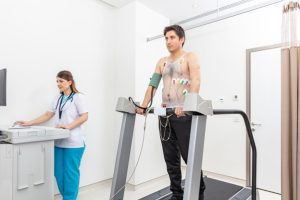
Figure 1: Treadmill stress test
Exercise Physiology
Treadmill stress testing causes a complex cascade of physiological changes, allowing for a thorough evaluation of cardiovascular function under exertional settings. The start of vigorous exercise stimulates the sympathetic nervous system while inhibiting parasympathetic tone. This autonomic shift causes an increase in heart rate, stroke volume, and cardiac output, which are all necessary cardiovascular responses to meet the increased metabolic and oxygen demands, particularly those of the myocardium.
During exercise, coronary blood flow increases significantly to meet myocardial oxygen requirements. This vasodilatory response is mediated by a variety of mechanisms, including endothelial vasoactive chemical production, passive dilation caused by increased coronary perfusion pressure, and decreased vasoconstrictive effects of circulating catecholamines. These actions together improve myocardial perfusion and reduce the risk of ischaemia. Importantly, this hyperemic state is blunted in stenotic coronary vessels, allowing for the detection of functionally significant coronary artery disease during stress testing.
Systemically, exercise induces several hemodynamic adaptations. Sympathetic-driven vasoconstriction enhances alveolar ventilation and venous return. Concurrently, skeletal muscle perfusion rises significantly, contributing to a decrease in total peripheral resistance. These responses cause an increase in systolic blood pressure (SBP), mean arterial pressure (MAP), and pulse pressure, while diastolic blood pressure (DBP) typically remains stable, increases slightly, or decreases modestly, usually within a 10 mm Hg range. The extent of these alterations is influenced by factors such as physical activity intensity, active muscle volume, and personal fitness levels.
In the early stages of exercise, increased cardiac output is primarily caused by increased stroke volume via the Frank-Starling mechanism and increased myocardial contractility. As exercise progresses, additional increases in cardiac output are mostly caused by an increased heart rate. At peak exertion, elevated plasma concentrations of norepinephrine and epinephrine enhance ventricular contractility and support blood flow redistribution, maintaining vasoconstriction in non-essential vascular regions while preserving perfusion to vital organs like the heart and brain.
Following the cessation of exercise, hemodynamic variables generally normalize within minutes. This recovery phase is primarily governed by the reactivation of parasympathetic tone, a process more efficient in physically conditioned individuals and attenuated in patients with chronic heart failure. Additionally, strenuous or prolonged exertion may result in an oxygen deficit, necessitating an elevated oxygen uptake during the recovery phase to repay the accumulated “oxygen debt.”
Indications
Treadmill stress testing is an important diagnostic and prognostic tool in cardiovascular medicine, primarily used in the evaluation of suspected coronary artery disease (CAD). It is particularly indicated in patients who are able to perform physical activity and who have a normal or near-normal resting electrocardiogram (ECG). The test assesses the heart’s reaction to graded exercise, gives useful information about myocardial perfusion, exercise tolerance, arrhythmic tendencies, and haemodynamic responses under stress.
Treadmill stress testing serves multiple clinical applications across a broad range of cardiovascular conditions. It is primarily indicated for diagnosing myocardial ischemia and coronary artery disease (CAD) in patients with symptoms like chest pain, dyspnea, or fatigue, provided they have a normal or near-normal resting ECG and are physically able to exercise.
In low-risk individuals presenting with acute chest pain but excluded for acute coronary syndrome (ACS), it helps to detect underlying obstructive disease. Following recent ACS without coronary angiography or incomplete revascularization, treadmill testing provides critical information on residual ischemia and functional status.
It is also useful for reassessing known CAD patients with worsening symptoms and for long-term surveillance following coronary revascularization specifically, five years after coronary artery bypass grafting (CABG) or within two years post-percutaneous coronary intervention (PCI) to detect silent ischemia or restenosis.
Treadmill testing helps to evaluate the functional capacity and hemodynamic response in valvular heart disease, identifying chronotropic incompetence and exercise-induced arrhythmias, and assessing functional capacity, ischemic burden, and prognosis in newly diagnosed heart failure or cardiomyopathy.
Contraindications
Absolute contraindications: These include a recent acute myocardial infarction (within the preceding 2–3 days), unstable angina that has not been medically stabilized, and uncontrolled arrhythmias that result in symptoms or compromise hemodynamic stability.
Additional absolute contraindications involve symptomatic severe aortic stenosis, decompensated heart failure, acute pulmonary embolism or infarction, severe pulmonary hypertension, and active inflammatory cardiac conditions like myocarditis, pericarditis, endocarditis, or acute aortic dissection.
Relative contraindications: These include high-grade atrioventricular (AV) conduction blocks, markedly elevated blood pressure (systolic >200 mmHg or diastolic >110 mmHg), and an inability to perform sufficient exercise due to severe obesity or physical or mental limitations.
Additional contraindications include significant electrolyte imbalances, known left main coronary artery disease, moderate valvular stenosis, bradyarrhythmias or tachyarrhythmias, hypertrophic cardiomyopathy, and any condition that restricts effective physical exertion.
Outcomes
While stress testing does not detect all individuals at high cardiovascular risk, the need for pharmacologic stress testing due to an inability to perform treadmill exercise has been identified as a strong independent predictor of mortality in patients with suspected CAD. Patients undergoing pharmacologic stress in the context of stress-rest single-photon emission computed tomography (SPECT) myocardial perfusion imaging (MPI) exhibit higher mortality across all body mass index (BMI) categories compared to those who complete exercise-based stress tests.
A 2022 systematic review and meta-analysis by Bingel et al., which included 140 study and 7,248 exercise tests examined hemodynamic responses in patients with both preserved and reduced ejection fraction heart failure during physiologic and pharmacologic stress testing. The findings showed that high-intensity stress tests produced greater absolute increases in heart rate, with age being a contributing factor. However, no significant differences were observed in heart rate response when comparing moderate- versus light-intensity exercise or pharmacologic versus dynamic stress testing.
Both pharmacologic and high-intensity stress protocols were associated with greater increases in ejection fraction, but there was no observed correlation between the type or intensity of stress and changes in stroke volume or cardiac output.
Equipment
Treadmill: A motorized treadmill specifically designed for exercise stress testing. It should allow for adjustable speed and incline to gradually increase the workload on the patient.
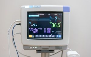
ECG Machine: A 12-lead ECG machine to record the heart’s electrical activity during exercise. This is crucial for detecting any abnormalities or changes in heart rhythm.
Figure 2: ECG machine
Electrodes: Tt is a small, flat, adhesive patches placed on the patient’s chest to connect them to the ECG machine.
Blood Pressure Monitor: A device to regularly measure blood pressure throughout the test.
Additional Considerations:
Crash Cart: A cart stocked with emergency medications, oxygen, and a defibrillator in case of any adverse events during the test.
Wireless ECG Acquisition Unit: Some modern systems use wireless ECG transmitters and receivers for greater patient comfort and freedom of movement.
Patient Preparation
To ensure accurate results and patient safety during treadmill stress testing, patients should arrive at least 15 minutes early and manage their medications properly.
Beta-blockers should not be taken 24 hours before the test, while other prescribed medications can be taken at their regular times. A light meal is allowed on the day, and patients should be well-hydrated.
Avoid coffee, tobacco, and nicotine-containing items might affect heart rate and blood pressure, thereby changing test results.
Clothing should be comfortable and appropriate for physical exercise, with males wearing loose-fitting shorts, jogging trousers, or athletic shoes and women in loose slacks or sports pants.
Patients should not use lotions, powders, or oils on the day of the test.
Patient Position
During a treadmill stress test, the patient begins in an upright, standing position on the treadmill.
Technique
Step 1: Pre-Test Preparation
The procedure begins with informed consent and a brief explanation of the test. A trained technologist or healthcare provider obtains a focused medical history, including medications, symptoms (especially chest pain or dyspnea), and prior cardiac events.
The technologist ensures the patient has adhered to pre-test instructions like avoiding beta-blockers, fasting, hydration, and wearing appropriate clothing.
Step 2: Electrode Placement and Baseline Measurements Adhesive ECG electrodes are applied to the chest after cleaning the skin with alcohol and shaving if needed. Mild abrasion may be used to reduce skin resistance and optimize signal quality. Electrodes are connected to the ECG machine for continuous monitoring. Baseline measurements include a resting 12-lead ECG, heart rate, and blood pressure, obtained in both supine and standing positions, as body posture can influence QRS and T-wave intervals.
Step 3: Evaluation of Resting ECG
Before initiating treadmill stress testing, a thorough assessment of the baseline ECG is essential, as certain abnormalities can interfere with accurate interpretation, especially for detecting ischemia. These interfering patterns may include ST-segment deviations (≥1 mm), left bundle branch block, paced rhythms, hypertrophy of either ventricle, pre-excitation syndromes like Wolff-Parkinson-White (WPW), strain-related T wave inversions, conduction delays, and drug-induced changes in ST-T segments. In such cases, combining the stress test with an imaging modality is recommended to enhance diagnostic accuracy. Overall, treadmill stress testing especially without imaging is more reliable in ruling out coronary artery disease (CAD) than confirming it.
Step 4: Initiation of Exercise Protocol
If the resting ECG is normal, the patient is placed on the treadmill. The patient begins walking on a treadmill. The choice of protocol depends on the individual’s exercise capacity:
Standard Bruce Protocol
Stage 1: 1.7 mph at 10% incline (~5 Metabolic Equivalent of Task [METs]) for 3 minutes.
Stage 2: 2.5 mph at 12% incline (~7 METs) for 3 minutes.
Stage 3: 3.4 mph at 14% incline (~9 METs) for 3 minutes.
Each subsequent stage increases speed and incline significantly.
Exercise capacity is reported in METs with 1 MET defined as 3.5 mL O₂/min/kg body weight.
Modified Bruce Protocol (commonly used in elderly or cardiac patients):
Warm-up Stage 1: 1.7 mph at 0% incline (~2 METs) for 3 minutes.
Warm-up Stage 2: 1.7 mph at 5% incline (~3 METs) for 3 minutes.
The protocol then continues with standard Bruce stages.
Step 4: Monitoring During Test
Continuous 12-lead ECG monitoring is performed throughout the test to detect ischemic changes, arrhythmias, or conduction abnormalities. Blood pressure is checked at each stage.
Patients are questioned for symptoms: chest pain, shortness of breath, fatigue, dizziness, or palpitations. The technologist monitors for onset of angina, dizziness, syncope, or extreme fatigue, abnormal ECG findings such as ST-segment depression or elevation and significant blood pressure changes or arrhythmias.
The patient is encouraged to walk as long as possible to achieve maximal effort unless symptoms or diagnostic endpoints require earlier termination.
Step 5: Post-Test Recovery
The treadmill is stopped, and the patient is asked to lie down or sit quietly. ECG, heart rate, and blood pressure are monitored every minute for 3–5 minutes or until vital signs return to baseline. Delayed symptoms such as chest discomfort or hypotension are also assessed.
Step 7: Data Review and Reporting
The ECG tracings, heart rate response, exercise duration, and symptoms are analyzed. The total METs achieved and any ST-segment changes are interpreted by a cardiologist.
Criteria for Termination of Test
The American College of Cardiology (ACC) and the American Heart Association (AHA) have defined certain conditions under which the test should be discontinued.
Absolute criteria for termination include a significant drop in systolic blood pressure (more than 10 mmHg) when accompanied by ischemic signs, moderate to severe angina, worsening neurological symptoms like dizziness, loss of coordination, or near-syncope, clinical signs of poor circulation like cyanosis or pallor, technical problems in tracking ECG or blood pressure, the patient’s request to stop, sustained episodes of ventricular tachycardia, and ST-segment elevations exceeding 1 mm in non-Q wave leads (excluding leads V1 or aVR).
Relative criteria for termination of the test are more discretionary and based on clinical judgment. These include a similar blood pressure drop in the absence of ischemia, excessive horizontal or downsloping ST depression greater than 2 mm, abnormal QRS changes or marked axis shifts, arrhythmias like supraventricular tachycardia, frequent or multifocal premature ventricular contractions, bradyarrhythmias, or varying degrees of heart block. Other signs include increasing fatigue, leg cramping, shortness of breath, wheezing, worsening chest discomfort, new conduction blocks indistinguishable from ventricular tachycardia, and extreme elevations in blood pressure (systolic ≥250 mmHg or diastolic >115 mmHg).
Treadmill Stress Test Interpretation
The interpretation of a treadmill stress test should provide a comprehensive evaluation of the patient’s exercise tolerance, clinical symptoms, hemodynamic response, and ECG changes. Notably, the emergence of chest pain characteristic of angina especially if it necessitates halting the test is clinically significant. Key visual indicators of a positive test include ST-segment deviations measured from the J point (the junction where the QRS complex ends and the ST segment begins) and the ST80 point (80 milliseconds after the J point). A depression of the ST segment by 0.1 mV (1 mm) or more, combined with a slope between ±1 mV/sec over three consecutive beats, is suggestive of myocardial ischemia.
However, not all ST-segment depressions indicate coronary artery disease (CAD). Several non-ischemic conditions can mimic this finding, including severe hypertension, aortic stenosis, cardiomyopathy, anemia, hypokalemia, hypoxia, digitalis use, abrupt strenuous activity, glucose intake, left ventricular hypertrophy, hyperventilation, mitral valve prolapse, conduction abnormalities like WPW syndrome, and severe volume overload or tachyarrhythmias.
A normal stress test reflects a physiological rise in heart rate and blood pressure without ischemic ECG changes or arrhythmias. Conversely, an inadequate rise or a drop in blood pressure during exertion, especially with signs of ischemia has negative prognostic implications. The presence of angina or more than 2 mm of ST depression before completing Stage 2 of the Bruce protocol, or ST changes that persist longer than five minutes into recovery, suggest more severe and high-risk ischemia.
Stress test outcomes are generally categorized as positive, negative, equivocal, or uninterpretable particularly when confounding factors like inadequate heart rate response affect results.
The Duke Treadmill Score (DTS) helps in quantifying ischemic risk based on exercise duration, ST-segment changes, and the presence or absence of angina during exertion. DTS scores range from +15 (low risk) to -25 (high risk). A score ≥5 denotes low risk with a 5-year survival of approximately 97%, while a score ≤-11 signifies high risk with about 65% 5-year survival. Intermediate scores (between -10 and +4) reflect a 90% 5-year survival and typically warrant further evaluation using imaging-based modalities.
Complications
One of the most frequent complications involves cardiac arrhythmias like premature atrial or ventricular contractions, supraventricular tachycardia, atrial fibrillation or even ventricular tachycardia with rare cases of sustained ventricular arrhythmias necessitating immediate intervention.
Physical exertion can cause myocardial ischaemia or infarction, especially in people with severe or undetected coronary artery disease, sometimes leading to an abrupt myocardial infarction.
Hypotension or syncope can develop as a result of reduced cardiac output or reflex vasodilation, particularly in those with obstructive heart diseases. Conversely, some patients may exhibit a hypertensive response to exercise, marked by a dramatic rise in blood pressure (systolic ≥250 mmHg or diastolic >115 mmHg), increasing the risk for complications like aortic dissection or stroke.
Exercise-induced angina is another possible symptom that can signal critical coronary obstruction. Additionally, musculoskeletal injuries like falls or muscle strains may occurs particularly in elderly or mobility-impaired patients.
Individuals with pulmonary conditions may experience respiratory distress or bronchospasm, requiring prompt cessation of the test. Technical difficulties, including faulty ECG or blood pressure readings can compromise monitoring accuracy, while psychological reactions like anxiety or panic may mimic cardiac symptoms and lead to early termination.
heart’s response under controlled conditions. It entails continuous electrocardiography (ECG) and blood pressure monitoring while the patient exercises on a treadmill using standardised procedures. This type of exercise testing is largely used to quantify functional capacity, evaluate cardiovascular prognosis, predict the likelihood and severity of coronary artery disease (CAD), and assess the efficacy of therapeutic therapies.
Exercise stress testing, particularly when combined with ECG, remains a cost-effective, accessible, and dependable approach for identifying and controlling cardiovascular disease. Other techniques like metabolic gas analysis and imaging may be added in selected patients to enhance diagnostic accuracy particularly in those with intermediate or prior risk of cardiovascular events.
Figure 1: Treadmill stress test
Exercise Physiology
Treadmill stress testing causes a complex cascade of physiological changes, allowing for a thorough evaluation of cardiovascular function under exertional settings. The start of vigorous exercise stimulates the sympathetic nervous system while inhibiting parasympathetic tone. This autonomic shift causes an increase in heart rate, stroke volume, and cardiac output, which are all necessary cardiovascular responses to meet the increased metabolic and oxygen demands, particularly those of the myocardium.
During exercise, coronary blood flow increases significantly to meet myocardial oxygen requirements. This vasodilatory response is mediated by a variety of mechanisms, including endothelial vasoactive chemical production, passive dilation caused by increased coronary perfusion pressure, and decreased vasoconstrictive effects of circulating catecholamines. These actions together improve myocardial perfusion and reduce the risk of ischaemia. Importantly, this hyperemic state is blunted in stenotic coronary vessels, allowing for the detection of functionally significant coronary artery disease during stress testing.
Systemically, exercise induces several hemodynamic adaptations. Sympathetic-driven vasoconstriction enhances alveolar ventilation and venous return. Concurrently, skeletal muscle perfusion rises significantly, contributing to a decrease in total peripheral resistance. These responses cause an increase in systolic blood pressure (SBP), mean arterial pressure (MAP), and pulse pressure, while diastolic blood pressure (DBP) typically remains stable, increases slightly, or decreases modestly, usually within a 10 mm Hg range. The extent of these alterations is influenced by factors such as physical activity intensity, active muscle volume, and personal fitness levels.
In the early stages of exercise, increased cardiac output is primarily caused by increased stroke volume via the Frank-Starling mechanism and increased myocardial contractility. As exercise progresses, additional increases in cardiac output are mostly caused by an increased heart rate. At peak exertion, elevated plasma concentrations of norepinephrine and epinephrine enhance ventricular contractility and support blood flow redistribution, maintaining vasoconstriction in non-essential vascular regions while preserving perfusion to vital organs like the heart and brain.
Following the cessation of exercise, hemodynamic variables generally normalize within minutes. This recovery phase is primarily governed by the reactivation of parasympathetic tone, a process more efficient in physically conditioned individuals and attenuated in patients with chronic heart failure. Additionally, strenuous or prolonged exertion may result in an oxygen deficit, necessitating an elevated oxygen uptake during the recovery phase to repay the accumulated “oxygen debt.”
Treadmill stress testing is an important diagnostic and prognostic tool in cardiovascular medicine, primarily used in the evaluation of suspected coronary artery disease (CAD). It is particularly indicated in patients who are able to perform physical activity and who have a normal or near-normal resting electrocardiogram (ECG). The test assesses the heart’s reaction to graded exercise, gives useful information about myocardial perfusion, exercise tolerance, arrhythmic tendencies, and haemodynamic responses under stress.
Treadmill stress testing serves multiple clinical applications across a broad range of cardiovascular conditions. It is primarily indicated for diagnosing myocardial ischemia and coronary artery disease (CAD) in patients with symptoms like chest pain, dyspnea, or fatigue, provided they have a normal or near-normal resting ECG and are physically able to exercise.
In low-risk individuals presenting with acute chest pain but excluded for acute coronary syndrome (ACS), it helps to detect underlying obstructive disease. Following recent ACS without coronary angiography or incomplete revascularization, treadmill testing provides critical information on residual ischemia and functional status.
It is also useful for reassessing known CAD patients with worsening symptoms and for long-term surveillance following coronary revascularization specifically, five years after coronary artery bypass grafting (CABG) or within two years post-percutaneous coronary intervention (PCI) to detect silent ischemia or restenosis.
Treadmill testing helps to evaluate the functional capacity and hemodynamic response in valvular heart disease, identifying chronotropic incompetence and exercise-induced arrhythmias, and assessing functional capacity, ischemic burden, and prognosis in newly diagnosed heart failure or cardiomyopathy.
Absolute contraindications: These include a recent acute myocardial infarction (within the preceding 2–3 days), unstable angina that has not been medically stabilized, and uncontrolled arrhythmias that result in symptoms or compromise hemodynamic stability.
Additional absolute contraindications involve symptomatic severe aortic stenosis, decompensated heart failure, acute pulmonary embolism or infarction, severe pulmonary hypertension, and active inflammatory cardiac conditions like myocarditis, pericarditis, endocarditis, or acute aortic dissection.
Relative contraindications: These include high-grade atrioventricular (AV) conduction blocks, markedly elevated blood pressure (systolic >200 mmHg or diastolic >110 mmHg), and an inability to perform sufficient exercise due to severe obesity or physical or mental limitations.
Additional contraindications include significant electrolyte imbalances, known left main coronary artery disease, moderate valvular stenosis, bradyarrhythmias or tachyarrhythmias, hypertrophic cardiomyopathy, and any condition that restricts effective physical exertion.
While stress testing does not detect all individuals at high cardiovascular risk, the need for pharmacologic stress testing due to an inability to perform treadmill exercise has been identified as a strong independent predictor of mortality in patients with suspected CAD. Patients undergoing pharmacologic stress in the context of stress-rest single-photon emission computed tomography (SPECT) myocardial perfusion imaging (MPI) exhibit higher mortality across all body mass index (BMI) categories compared to those who complete exercise-based stress tests.
A 2022 systematic review and meta-analysis by Bingel et al., which included 140 study and 7,248 exercise tests examined hemodynamic responses in patients with both preserved and reduced ejection fraction heart failure during physiologic and pharmacologic stress testing. The findings showed that high-intensity stress tests produced greater absolute increases in heart rate, with age being a contributing factor. However, no significant differences were observed in heart rate response when comparing moderate- versus light-intensity exercise or pharmacologic versus dynamic stress testing.
Both pharmacologic and high-intensity stress protocols were associated with greater increases in ejection fraction, but there was no observed correlation between the type or intensity of stress and changes in stroke volume or cardiac output.
Treadmill: A motorized treadmill specifically designed for exercise stress testing. It should allow for adjustable speed and incline to gradually increase the workload on the patient.
ECG Machine: A 12-lead ECG machine to record the heart’s electrical activity during exercise. This is crucial for detecting any abnormalities or changes in heart rhythm.
Figure 2: ECG machine
Electrodes: Tt is a small, flat, adhesive patches placed on the patient’s chest to connect them to the ECG machine.
Blood Pressure Monitor: A device to regularly measure blood pressure throughout the test.
Additional Considerations:
Crash Cart: A cart stocked with emergency medications, oxygen, and a defibrillator in case of any adverse events during the test.
Wireless ECG Acquisition Unit: Some modern systems use wireless ECG transmitters and receivers for greater patient comfort and freedom of movement.
To ensure accurate results and patient safety during treadmill stress testing, patients should arrive at least 15 minutes early and manage their medications properly.
Beta-blockers should not be taken 24 hours before the test, while other prescribed medications can be taken at their regular times. A light meal is allowed on the day, and patients should be well-hydrated.
Avoid coffee, tobacco, and nicotine-containing items might affect heart rate and blood pressure, thereby changing test results.
Clothing should be comfortable and appropriate for physical exercise, with males wearing loose-fitting shorts, jogging trousers, or athletic shoes and women in loose slacks or sports pants.
Patients should not use lotions, powders, or oils on the day of the test.
During a treadmill stress test, the patient begins in an upright, standing position on the treadmill.
Step 1: Pre-Test Preparation
The procedure begins with informed consent and a brief explanation of the test. A trained technologist or healthcare provider obtains a focused medical history, including medications, symptoms (especially chest pain or dyspnea), and prior cardiac events.
The technologist ensures the patient has adhered to pre-test instructions like avoiding beta-blockers, fasting, hydration, and wearing appropriate clothing.
Step 2: Electrode Placement and Baseline Measurements Adhesive ECG electrodes are applied to the chest after cleaning the skin with alcohol and shaving if needed. Mild abrasion may be used to reduce skin resistance and optimize signal quality. Electrodes are connected to the ECG machine for continuous monitoring. Baseline measurements include a resting 12-lead ECG, heart rate, and blood pressure, obtained in both supine and standing positions, as body posture can influence QRS and T-wave intervals.
Step 3: Evaluation of Resting ECG
Before initiating treadmill stress testing, a thorough assessment of the baseline ECG is essential, as certain abnormalities can interfere with accurate interpretation, especially for detecting ischemia. These interfering patterns may include ST-segment deviations (≥1 mm), left bundle branch block, paced rhythms, hypertrophy of either ventricle, pre-excitation syndromes like Wolff-Parkinson-White (WPW), strain-related T wave inversions, conduction delays, and drug-induced changes in ST-T segments. In such cases, combining the stress test with an imaging modality is recommended to enhance diagnostic accuracy. Overall, treadmill stress testing especially without imaging is more reliable in ruling out coronary artery disease (CAD) than confirming it.
Step 4: Initiation of Exercise Protocol
If the resting ECG is normal, the patient is placed on the treadmill. The patient begins walking on a treadmill. The choice of protocol depends on the individual’s exercise capacity:
Stage 1: 1.7 mph at 10% incline (~5 Metabolic Equivalent of Task [METs]) for 3 minutes.
Stage 2: 2.5 mph at 12% incline (~7 METs) for 3 minutes.
Stage 3: 3.4 mph at 14% incline (~9 METs) for 3 minutes.
Each subsequent stage increases speed and incline significantly.
Exercise capacity is reported in METs with 1 MET defined as 3.5 mL O₂/min/kg body weight.
Modified Bruce Protocol (commonly used in elderly or cardiac patients):
Warm-up Stage 1: 1.7 mph at 0% incline (~2 METs) for 3 minutes.
Warm-up Stage 2: 1.7 mph at 5% incline (~3 METs) for 3 minutes.
The protocol then continues with standard Bruce stages.
Step 4: Monitoring During Test
Continuous 12-lead ECG monitoring is performed throughout the test to detect ischemic changes, arrhythmias, or conduction abnormalities. Blood pressure is checked at each stage.
Patients are questioned for symptoms: chest pain, shortness of breath, fatigue, dizziness, or palpitations. The technologist monitors for onset of angina, dizziness, syncope, or extreme fatigue, abnormal ECG findings such as ST-segment depression or elevation and significant blood pressure changes or arrhythmias.
The patient is encouraged to walk as long as possible to achieve maximal effort unless symptoms or diagnostic endpoints require earlier termination.
Step 5: Post-Test Recovery
The treadmill is stopped, and the patient is asked to lie down or sit quietly. ECG, heart rate, and blood pressure are monitored every minute for 3–5 minutes or until vital signs return to baseline. Delayed symptoms such as chest discomfort or hypotension are also assessed.
Step 7: Data Review and Reporting
The ECG tracings, heart rate response, exercise duration, and symptoms are analyzed. The total METs achieved and any ST-segment changes are interpreted by a cardiologist.
The American College of Cardiology (ACC) and the American Heart Association (AHA) have defined certain conditions under which the test should be discontinued.
Absolute criteria for termination include a significant drop in systolic blood pressure (more than 10 mmHg) when accompanied by ischemic signs, moderate to severe angina, worsening neurological symptoms like dizziness, loss of coordination, or near-syncope, clinical signs of poor circulation like cyanosis or pallor, technical problems in tracking ECG or blood pressure, the patient’s request to stop, sustained episodes of ventricular tachycardia, and ST-segment elevations exceeding 1 mm in non-Q wave leads (excluding leads V1 or aVR).
Relative criteria for termination of the test are more discretionary and based on clinical judgment. These include a similar blood pressure drop in the absence of ischemia, excessive horizontal or downsloping ST depression greater than 2 mm, abnormal QRS changes or marked axis shifts, arrhythmias like supraventricular tachycardia, frequent or multifocal premature ventricular contractions, bradyarrhythmias, or varying degrees of heart block. Other signs include increasing fatigue, leg cramping, shortness of breath, wheezing, worsening chest discomfort, new conduction blocks indistinguishable from ventricular tachycardia, and extreme elevations in blood pressure (systolic ≥250 mmHg or diastolic >115 mmHg).
The interpretation of a treadmill stress test should provide a comprehensive evaluation of the patient’s exercise tolerance, clinical symptoms, hemodynamic response, and ECG changes. Notably, the emergence of chest pain characteristic of angina especially if it necessitates halting the test is clinically significant. Key visual indicators of a positive test include ST-segment deviations measured from the J point (the junction where the QRS complex ends and the ST segment begins) and the ST80 point (80 milliseconds after the J point). A depression of the ST segment by 0.1 mV (1 mm) or more, combined with a slope between ±1 mV/sec over three consecutive beats, is suggestive of myocardial ischemia.
However, not all ST-segment depressions indicate coronary artery disease (CAD). Several non-ischemic conditions can mimic this finding, including severe hypertension, aortic stenosis, cardiomyopathy, anemia, hypokalemia, hypoxia, digitalis use, abrupt strenuous activity, glucose intake, left ventricular hypertrophy, hyperventilation, mitral valve prolapse, conduction abnormalities like WPW syndrome, and severe volume overload or tachyarrhythmias.
A normal stress test reflects a physiological rise in heart rate and blood pressure without ischemic ECG changes or arrhythmias. Conversely, an inadequate rise or a drop in blood pressure during exertion, especially with signs of ischemia has negative prognostic implications. The presence of angina or more than 2 mm of ST depression before completing Stage 2 of the Bruce protocol, or ST changes that persist longer than five minutes into recovery, suggest more severe and high-risk ischemia.
Stress test outcomes are generally categorized as positive, negative, equivocal, or uninterpretable particularly when confounding factors like inadequate heart rate response affect results.
The Duke Treadmill Score (DTS) helps in quantifying ischemic risk based on exercise duration, ST-segment changes, and the presence or absence of angina during exertion. DTS scores range from +15 (low risk) to -25 (high risk). A score ≥5 denotes low risk with a 5-year survival of approximately 97%, while a score ≤-11 signifies high risk with about 65% 5-year survival. Intermediate scores (between -10 and +4) reflect a 90% 5-year survival and typically warrant further evaluation using imaging-based modalities.
One of the most frequent complications involves cardiac arrhythmias like premature atrial or ventricular contractions, supraventricular tachycardia, atrial fibrillation or even ventricular tachycardia with rare cases of sustained ventricular arrhythmias necessitating immediate intervention.
Physical exertion can cause myocardial ischaemia or infarction, especially in people with severe or undetected coronary artery disease, sometimes leading to an abrupt myocardial infarction.
Hypotension or syncope can develop as a result of reduced cardiac output or reflex vasodilation, particularly in those with obstructive heart diseases. Conversely, some patients may exhibit a hypertensive response to exercise, marked by a dramatic rise in blood pressure (systolic ≥250 mmHg or diastolic >115 mmHg), increasing the risk for complications like aortic dissection or stroke.
Exercise-induced angina is another possible symptom that can signal critical coronary obstruction. Additionally, musculoskeletal injuries like falls or muscle strains may occurs particularly in elderly or mobility-impaired patients.
Individuals with pulmonary conditions may experience respiratory distress or bronchospasm, requiring prompt cessation of the test. Technical difficulties, including faulty ECG or blood pressure readings can compromise monitoring accuracy, while psychological reactions like anxiety or panic may mimic cardiac symptoms and lead to early termination.
Both our subscription plans include Free CME/CPD AMA PRA Category 1 credits.
Digital Certificate PDF
On course completion, you will receive a full-sized presentation quality digital certificate.
medtigo Simulation
A dynamic medical simulation platform designed to train healthcare professionals and students to effectively run code situations through an immersive hands-on experience in a live, interactive 3D environment.
medtigo Points
medtigo points is our unique point redemption system created to award users for interacting on our site. These points can be redeemed for special discounts on the medtigo marketplace as well as towards the membership cost itself.
Community Forum post/reply = 5 points
*Redemption of points can occur only through the medtigo marketplace, courses, or simulation system. Money will not be credited to your bank account. 10 points = $1.
All Your Certificates in One Place
When you have your licenses, certificates and CMEs in one place, it's easier to track your career growth. You can easily share these with hospitals as well, using your medtigo app.