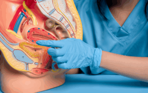
Uterine balloon tamponade is an option for medical treatment that is employed to control postpartum hemorrhage (PPH), a possible life-threatening situation happening after childbirth. In the 21st century, it is widely used as a safe and minimally invasive intervention that stands out from other more aggressive operations, thus enabling effective care of PPH, especially in resource-limited areas. Uterine balloon tamponade’s prime purpose is to bring pressure on the uterine walls that, in its turn, will compress the blood vessels and limit the bleeding. Thereby, through sterile fluid, a special balloon device is placed in the uterine cavity and filled to the correct level. The enhanced pressure which is attained by this action, enables hemorrhage control and blood clam formation that in turn avoids the loss of more blood. As a result, uterine tampon is now an essential feature of the broader PPH management strategy, which now includes uterotonic medications, surgical interventions, and other obstetric practices. Its continued availability and possibility of use in non-surgical areas make it a valuable tool where immediate surgical options are limited. Nonetheless, it is safe to say that proper training, infection control, patient comfort, and equipment availability are the four most important factors to guarantee that it is implemented efficiently.
Indications
Uterine balloon tamponade is a procedure that is carried out when a woman has postpartum hemorrhage (PPH) caused by the absence of conventional methods, such as massage, uterotonic drugs, or manual compression. Uterine balloon tamponade is a good alternative usage in which uterotonic medications fail to contract and control the bleeding, thus preventing excess bleeding. Uterine balloon tamponade is also used during trauma related to childbirth, placental abnormalities coagulopathy, postpartum hemorrhage during or after a cesarean section and intrauterine infections. Additionally, the utility of this method is even more in low-resource settings where surgeries can be difficult to access quickly. The patients’ overall health and morbidity conditions may also matter with regard to the decision as to whether to use uterine balloon tamponade. The overall health state of the patient and the importance of the situation solve the dilemma of whether to use uterine balloon tamponade or not. In conclusion, uterine balloon tamponade holds a lot of advantages in the treatment of PPH in different scenarios like uterine atony, failures of the uterotonic drugs, trauma, placental abnormalities, coagulopathy, postpartum hemorrhage during cesarean sections and intrauterine infections.
Contraindications
Uterine balloon tamponade is not recommended in the event of known or suspected uterine perforations, active uterine infections, unresponsive uterine atony, severe coagulopathy or bleeding disorders, uterine malignancy, allergies or sensitivities to balloon materials, severe pelvic adhesions, gestational trophoblastic disease, gestational age beyond term, or patient refusal or lack of informed consent. Uterine balloon tamponade may not have the desired effect in such cases, and in actuality its effectiveness may be restricted in postpartum hemorrhage cases going beyond the term of gestation. In such cases alternative interventions may be more favorable. Severe pelvic adhesions may interfere with the correct placement, and inflation of the balloon making its usage limit the desired effectiveness. Uterine balloon tamponade may be incapable of accomplishing the prevention of the destined postpartum hemorrhage in case of gestational trophoblastic disease, and its impact may be restricted in postpartum hemorrhage cases occurring after the term of gestation. mother needs to give her informed consent for the uterine balloon tamponade but she should be informed of the risks and benefits of the procedure. If a patient declines the operation or cannot give her permission, it should not be done.
Outcomes
Uterine balloon tamponade is the intervention for postpartum hemorrhage, which is a surgical procedure. It uses the force to contract the uterine walls, blood vessels, diminish blood flow, and therefore get hemostasis. The effect of that is the stabilization of the patient by the consumption of the excess blood and improving the functional and haemodynamic stability. If the tamponade is successful, a decrease in the number of blood transfusions required will be observed, thus minimizing blood loss and associated complications. It ends with the ultimate treatment, letting health care workers have preparation time for more definitive treatments. Uterine balloon tamponade yields positive outcomes for the mothers, saving lives and preventing serious complications connected with the uncontrolled blood loss. Thus, the risk of complications, for example, DIC, organ failure, and extensive surgical interventions are among these. Equally, it keeps the reproductive system working by averting severe hemorrhagic events.
Equipment
The balloon device is the significant and essential part in the tamponade of the uterus and the control of postpartum hemorrhage. It is connected to a catheter or tubing, which is filled with the balloon via sterile fluid. The choice of the fluid option is determined by the specific rules of each hospital and recommendations of each company. The medical equipment is also equipped with an inflation port or valve, which is responsible for the gradual filling of the sterile fluid. Some types of balloon devices are equipped with a special system that measures the pressure inside the uterus. A syringe or infusion pump will be required to instill the sterile fluid. A speculum serves the purpose of visualization and insertion of the silicone balloon to reach the cervix. Cervical dilators are among such instruments that are employed to the cervical dilations if required. The anesthesia can be administered topically to minimize the discomfort during balloon insertion. A fixation element (clamp/velcro strip/ribbon) is used to fix the catheter or tubing to the patient’s thigh or abdomen. Ultrasound, as well as balloon-guided procedure, can be included in the interventional room. Accessories that are free of germs like gloves, surgical drapes, and uninfected gauze are employed to aseptically execute the procedure.
Patient preparation
Upon the informed consent of the woman or legal representative, the uterine balloon tamponade procedure is carried out. The physical exam is performed only after the patient has been briefed and signed a consent form which gives permission to conduct the procedure, the procedure to be performed, and all the other relevant details. The patient’s position is usually in the lithotomy position so to make insertion of the tube and access easier to the perineum. To prevent infection, professionals create a sterile field and put on sterile gloves. They also keep the standard aseptic rules. This includes a speculum exam revealing the cervix and checking if it is dilated enough for the procedure. In case the sutures need to be performed, then the nursing staff involves using a balloon-tipped catheter and is guided by the doctors. The staff also ensures that each step is clearly understood by the patient and the explanations are clear and concise. Regular communication is needed since the patient should be informed about every step and the staff should be quick enough to deal with any discomfort or concerns. The very step includes coming up with a development plan involving the patient to communicate if they were in pain or distress during operation.
Monitoring and Follow-up
Continuous monitoring of vital signs is the keystone for an early hemodynamic stability alert. The first thing should be to check for the presence of uterine bleeding in order to see if the balloon is being effective. Pain management and comfort measurement are the key not only for recovering comfort and well-being but also for dealing with psychological health. The recorded volumes of fluid insertion and the pressure developed are necessary for assessing the procedure. The patient’s status is checked at the end of short clinical assessments, which involve peritonitis, adnexa enlargement, and signs of sepsis. Ultrasound imaging is the most used modality to verify the balloon location inside the uterine cavity, and sequential hemoglobin and hematocrit tests are approaches for monitoring blood volume, with transfusion being the treatment option if necessary. The management of coagulopathy and blood typing are two main laboratory tests that are usually performed. Patient communication in which a discussion takes place if required is the dominant piece of advice. The need for balloon continuous packing is regularly revised, and if hemostasis is effective and the patient is stable, the balloon can be removed. Uterine artery embolization or surgical procedures can be the final way of therapy. The follow-up plan is a quintessential requirement both to note the occurrence of recurrent bleeding or complication and to develop required interventions and support.
Uterine balloon tamponade
Step-1: Patient preparation: Inform the patient or their legal representative of the procedure and secure their consent. Explain the process in detail, its objectives, as well as the potential benefits and risks. Diagnose the patient’s medical history, allergies, and general health condition. Make sure the patient is lying in a lithotomic position to have a proper look at the perineum for the procedure.
Step-2: Sterile field set up: The operating room or delivery suite should be made into the sterile field properly. To start off with, make sure that all the necessary materials, such as sterile gloves, drapes, and instruments, are at hand. Stick to the proper standard aseptic techniques right through the operation during the procedure.
Step-3: Gather equipment: Check the preparation and functionality of the chosen uterine balloon tamponade device for the operation. These devices are usually the Bakri balloon, condom catheter balloon, or other commercially obtained products. Check if the catheter or tubing, the inflation port or valve, and other components are unharmed.
Step-4: Local anesthesia: Apply a pain blocker directly to the cervix or the low segment of the uterus to provide the patient with the most comfort possible during the examination. Profound anesthesia is quite necessary if cervical dilation is required.
Step-5: Cervical examination: Performing a speculum examination is the proper procedure to observe and reach the cervix. The position and condition of the cervix should be evaluated and in case of need, dilators or a balloon-tipped catheter may be used to go through cervical dilation.
Step-6: Balloon insertion: Pass the deflated balloon device through the cervix and into the uterine cavity as part of the introduction. Make sure that the insertion procedure is done gently to prevent trauma. The balloon has to be put in the middle of the uterine cavity to some extent, to provide a muscle-pulling effect characteristic of tamponade, and thus its tamponade effect is maximized.
Step-7: Balloon inflation: Attach the catheter or tubing to a syringe or infusion pump that is filled with sterile fluid, usually, normal saline, or sterile water. Begin to inflate the balloon slowly while ensuring that observed the volume and pressure is maintained. Adhere to the manufacturer’s specifications of the recommended inflation volume.
Step-8: Pressure monitoring (Optional): If the device is installed with a pressure monitoring system, use it to measure intrauterine pressure and the progress of labor. Measurement of pressure is critical in determining whether the tamponade was successfully done or whether adjustments are still needed.
Step-9: Securing the catheter: After the balloon has been pumped to the optimum dimension, tie the catheter or tubing to the thigh or abdomen of the patient. This safeguards the smooth sailing of the balloon, maintaining a uniform degree of tension in the uterine chamber.
Step-10: Ultrasound confirmation (Optional): When ultrasound imaging is ready, do a further ultrasound to see the position of the balloon inside the uterus chamber. Make sure the balloon does not affect the cervix or cause uterus disturbances.
Step-11: Regular clinical assessments: Conduct a series of clinical assessments, which is an abdominal examination and an evaluation of the size of the uterine. Observe for any signs of infection, abdominal tenderness, or any uterine balloon tamponade complications.
Step-12: Balloon duration assessment: Regularly monitor the patient’s response and clinical status to determine if the balloon tamponade intervention is still necessary. The balloon may be removed when the bleeding has been properly managed, and the patient becomes stable.
Step-13: Balloon removal: If the decision to remove the balloon has been made, first, the balloon must be carefully deflated through the sterile fluid extraction. The deflated balloon device should be pulled out gently and quietly to avoid causing damage.
Complications
Balloon tamponade can be compromised by many reasons, among which unintentional balloon displacement, balloon rupture, uterine perforation, infection, retained placental tissue, cervical laceration, vaginal bleeding, catheter-related complications, hematometra, coagulopathy, and hypovolemia are the main ones. The loss of the desired tamponade effect can be caused by the displacement of the balloon and the bleeding might not be stopped, which would lead to inadequate control. The balloon becoming too tight or having damage to the material may result in the balloon’s rupture which in turn can lead to more bleeding. The perforation of the uterus can potentially be the cause of bladder or the bowel damage. Infection may develop through the introduction of a foreign material during the insertion of a balloon. Catheter-related complications can result in the malfunctioning of a tamponade by cases like kinking, obstruction, or accidental dislodgment issues. Tamponade failure can lead to hematometra when blood is accumulated in the uterine cavity. Very severe coagulopathy may demand further interventions, and inattention to the proper resuscitation or the failure to deal with the root cause of the hemorrhage can result in the continuous low blood pressure.
Uterine balloon tamponade
Medication summary
Local anesthetic agents: Lidocaine or bupivacaine can be administered to the cervix before insertion of balloon to ease the discomfort during procedure.
Sterile fluid for balloon inflation: Typically, normal saline or sterile water may be used to inflate the balloon. The choice of the fluid depends on the recommendations of the manufacturer.
Antibiotics: Ampicillin and cefotaxime is often used as a prophylactic treatment to lower the risk of infection associated with the introduction of foreign material during balloon insertion.
Medications for pain management: These include the use of opiods such as fentanyl or morphine or other analgesics to overcome pain during the procedure.
Diuretics: Furosemide or chlorothiazide may be prescribed to facilitate the fluid mobilization after the procedure.
Antacids and H2 antagonists: These drugs may be used to treat gastrointestinal bleeding which is a major complication of uterine balloon tamponade.
Phenobarbital: This is an anticonvulsant medication used to prevent and control seizures associated with postpartum hemorrhage
Hemostatic medications (if needed): Hemostatic medications such as tranexamic acid or recombinant factor VIIa may be used in cases of persistent bleeding or coagulopathy to promote clotting of blood and enhance hemostasis.
Uterine balloon tamponade is an option for medical treatment that is employed to control postpartum hemorrhage (PPH), a possible life-threatening situation happening after childbirth. In the 21st century, it is widely used as a safe and minimally invasive intervention that stands out from other more aggressive operations, thus enabling effective care of PPH, especially in resource-limited areas. Uterine balloon tamponade’s prime purpose is to bring pressure on the uterine walls that, in its turn, will compress the blood vessels and limit the bleeding. Thereby, through sterile fluid, a special balloon device is placed in the uterine cavity and filled to the correct level. The enhanced pressure which is attained by this action, enables hemorrhage control and blood clam formation that in turn avoids the loss of more blood. As a result, uterine tampon is now an essential feature of the broader PPH management strategy, which now includes uterotonic medications, surgical interventions, and other obstetric practices. Its continued availability and possibility of use in non-surgical areas make it a valuable tool where immediate surgical options are limited. Nonetheless, it is safe to say that proper training, infection control, patient comfort, and equipment availability are the four most important factors to guarantee that it is implemented efficiently.
Uterine balloon tamponade is a procedure that is carried out when a woman has postpartum hemorrhage (PPH) caused by the absence of conventional methods, such as massage, uterotonic drugs, or manual compression. Uterine balloon tamponade is a good alternative usage in which uterotonic medications fail to contract and control the bleeding, thus preventing excess bleeding. Uterine balloon tamponade is also used during trauma related to childbirth, placental abnormalities coagulopathy, postpartum hemorrhage during or after a cesarean section and intrauterine infections. Additionally, the utility of this method is even more in low-resource settings where surgeries can be difficult to access quickly. The patients’ overall health and morbidity conditions may also matter with regard to the decision as to whether to use uterine balloon tamponade. The overall health state of the patient and the importance of the situation solve the dilemma of whether to use uterine balloon tamponade or not. In conclusion, uterine balloon tamponade holds a lot of advantages in the treatment of PPH in different scenarios like uterine atony, failures of the uterotonic drugs, trauma, placental abnormalities, coagulopathy, postpartum hemorrhage during cesarean sections and intrauterine infections.
Uterine balloon tamponade is not recommended in the event of known or suspected uterine perforations, active uterine infections, unresponsive uterine atony, severe coagulopathy or bleeding disorders, uterine malignancy, allergies or sensitivities to balloon materials, severe pelvic adhesions, gestational trophoblastic disease, gestational age beyond term, or patient refusal or lack of informed consent. Uterine balloon tamponade may not have the desired effect in such cases, and in actuality its effectiveness may be restricted in postpartum hemorrhage cases going beyond the term of gestation. In such cases alternative interventions may be more favorable. Severe pelvic adhesions may interfere with the correct placement, and inflation of the balloon making its usage limit the desired effectiveness. Uterine balloon tamponade may be incapable of accomplishing the prevention of the destined postpartum hemorrhage in case of gestational trophoblastic disease, and its impact may be restricted in postpartum hemorrhage cases occurring after the term of gestation. mother needs to give her informed consent for the uterine balloon tamponade but she should be informed of the risks and benefits of the procedure. If a patient declines the operation or cannot give her permission, it should not be done.
Uterine balloon tamponade is the intervention for postpartum hemorrhage, which is a surgical procedure. It uses the force to contract the uterine walls, blood vessels, diminish blood flow, and therefore get hemostasis. The effect of that is the stabilization of the patient by the consumption of the excess blood and improving the functional and haemodynamic stability. If the tamponade is successful, a decrease in the number of blood transfusions required will be observed, thus minimizing blood loss and associated complications. It ends with the ultimate treatment, letting health care workers have preparation time for more definitive treatments. Uterine balloon tamponade yields positive outcomes for the mothers, saving lives and preventing serious complications connected with the uncontrolled blood loss. Thus, the risk of complications, for example, DIC, organ failure, and extensive surgical interventions are among these. Equally, it keeps the reproductive system working by averting severe hemorrhagic events.
The balloon device is the significant and essential part in the tamponade of the uterus and the control of postpartum hemorrhage. It is connected to a catheter or tubing, which is filled with the balloon via sterile fluid. The choice of the fluid option is determined by the specific rules of each hospital and recommendations of each company. The medical equipment is also equipped with an inflation port or valve, which is responsible for the gradual filling of the sterile fluid. Some types of balloon devices are equipped with a special system that measures the pressure inside the uterus. A syringe or infusion pump will be required to instill the sterile fluid. A speculum serves the purpose of visualization and insertion of the silicone balloon to reach the cervix. Cervical dilators are among such instruments that are employed to the cervical dilations if required. The anesthesia can be administered topically to minimize the discomfort during balloon insertion. A fixation element (clamp/velcro strip/ribbon) is used to fix the catheter or tubing to the patient’s thigh or abdomen. Ultrasound, as well as balloon-guided procedure, can be included in the interventional room. Accessories that are free of germs like gloves, surgical drapes, and uninfected gauze are employed to aseptically execute the procedure.
Upon the informed consent of the woman or legal representative, the uterine balloon tamponade procedure is carried out. The physical exam is performed only after the patient has been briefed and signed a consent form which gives permission to conduct the procedure, the procedure to be performed, and all the other relevant details. The patient’s position is usually in the lithotomy position so to make insertion of the tube and access easier to the perineum. To prevent infection, professionals create a sterile field and put on sterile gloves. They also keep the standard aseptic rules. This includes a speculum exam revealing the cervix and checking if it is dilated enough for the procedure. In case the sutures need to be performed, then the nursing staff involves using a balloon-tipped catheter and is guided by the doctors. The staff also ensures that each step is clearly understood by the patient and the explanations are clear and concise. Regular communication is needed since the patient should be informed about every step and the staff should be quick enough to deal with any discomfort or concerns. The very step includes coming up with a development plan involving the patient to communicate if they were in pain or distress during operation.
Continuous monitoring of vital signs is the keystone for an early hemodynamic stability alert. The first thing should be to check for the presence of uterine bleeding in order to see if the balloon is being effective. Pain management and comfort measurement are the key not only for recovering comfort and well-being but also for dealing with psychological health. The recorded volumes of fluid insertion and the pressure developed are necessary for assessing the procedure. The patient’s status is checked at the end of short clinical assessments, which involve peritonitis, adnexa enlargement, and signs of sepsis. Ultrasound imaging is the most used modality to verify the balloon location inside the uterine cavity, and sequential hemoglobin and hematocrit tests are approaches for monitoring blood volume, with transfusion being the treatment option if necessary. The management of coagulopathy and blood typing are two main laboratory tests that are usually performed. Patient communication in which a discussion takes place if required is the dominant piece of advice. The need for balloon continuous packing is regularly revised, and if hemostasis is effective and the patient is stable, the balloon can be removed. Uterine artery embolization or surgical procedures can be the final way of therapy. The follow-up plan is a quintessential requirement both to note the occurrence of recurrent bleeding or complication and to develop required interventions and support.
Step-1: Patient preparation: Inform the patient or their legal representative of the procedure and secure their consent. Explain the process in detail, its objectives, as well as the potential benefits and risks. Diagnose the patient’s medical history, allergies, and general health condition. Make sure the patient is lying in a lithotomic position to have a proper look at the perineum for the procedure.
Step-2: Sterile field set up: The operating room or delivery suite should be made into the sterile field properly. To start off with, make sure that all the necessary materials, such as sterile gloves, drapes, and instruments, are at hand. Stick to the proper standard aseptic techniques right through the operation during the procedure.
Step-3: Gather equipment: Check the preparation and functionality of the chosen uterine balloon tamponade device for the operation. These devices are usually the Bakri balloon, condom catheter balloon, or other commercially obtained products. Check if the catheter or tubing, the inflation port or valve, and other components are unharmed.
Step-4: Local anesthesia: Apply a pain blocker directly to the cervix or the low segment of the uterus to provide the patient with the most comfort possible during the examination. Profound anesthesia is quite necessary if cervical dilation is required.
Step-5: Cervical examination: Performing a speculum examination is the proper procedure to observe and reach the cervix. The position and condition of the cervix should be evaluated and in case of need, dilators or a balloon-tipped catheter may be used to go through cervical dilation.
Step-6: Balloon insertion: Pass the deflated balloon device through the cervix and into the uterine cavity as part of the introduction. Make sure that the insertion procedure is done gently to prevent trauma. The balloon has to be put in the middle of the uterine cavity to some extent, to provide a muscle-pulling effect characteristic of tamponade, and thus its tamponade effect is maximized.
Step-7: Balloon inflation: Attach the catheter or tubing to a syringe or infusion pump that is filled with sterile fluid, usually, normal saline, or sterile water. Begin to inflate the balloon slowly while ensuring that observed the volume and pressure is maintained. Adhere to the manufacturer’s specifications of the recommended inflation volume.
Step-8: Pressure monitoring (Optional): If the device is installed with a pressure monitoring system, use it to measure intrauterine pressure and the progress of labor. Measurement of pressure is critical in determining whether the tamponade was successfully done or whether adjustments are still needed.
Step-9: Securing the catheter: After the balloon has been pumped to the optimum dimension, tie the catheter or tubing to the thigh or abdomen of the patient. This safeguards the smooth sailing of the balloon, maintaining a uniform degree of tension in the uterine chamber.
Step-10: Ultrasound confirmation (Optional): When ultrasound imaging is ready, do a further ultrasound to see the position of the balloon inside the uterus chamber. Make sure the balloon does not affect the cervix or cause uterus disturbances.
Step-11: Regular clinical assessments: Conduct a series of clinical assessments, which is an abdominal examination and an evaluation of the size of the uterine. Observe for any signs of infection, abdominal tenderness, or any uterine balloon tamponade complications.
Step-12: Balloon duration assessment: Regularly monitor the patient’s response and clinical status to determine if the balloon tamponade intervention is still necessary. The balloon may be removed when the bleeding has been properly managed, and the patient becomes stable.
Step-13: Balloon removal: If the decision to remove the balloon has been made, first, the balloon must be carefully deflated through the sterile fluid extraction. The deflated balloon device should be pulled out gently and quietly to avoid causing damage.
Balloon tamponade can be compromised by many reasons, among which unintentional balloon displacement, balloon rupture, uterine perforation, infection, retained placental tissue, cervical laceration, vaginal bleeding, catheter-related complications, hematometra, coagulopathy, and hypovolemia are the main ones. The loss of the desired tamponade effect can be caused by the displacement of the balloon and the bleeding might not be stopped, which would lead to inadequate control. The balloon becoming too tight or having damage to the material may result in the balloon’s rupture which in turn can lead to more bleeding. The perforation of the uterus can potentially be the cause of bladder or the bowel damage. Infection may develop through the introduction of a foreign material during the insertion of a balloon. Catheter-related complications can result in the malfunctioning of a tamponade by cases like kinking, obstruction, or accidental dislodgment issues. Tamponade failure can lead to hematometra when blood is accumulated in the uterine cavity. Very severe coagulopathy may demand further interventions, and inattention to the proper resuscitation or the failure to deal with the root cause of the hemorrhage can result in the continuous low blood pressure.
Uterine balloon tamponade
Local anesthetic agents: Lidocaine or bupivacaine can be administered to the cervix before insertion of balloon to ease the discomfort during procedure.
Sterile fluid for balloon inflation: Typically, normal saline or sterile water may be used to inflate the balloon. The choice of the fluid depends on the recommendations of the manufacturer.
Antibiotics: Ampicillin and cefotaxime is often used as a prophylactic treatment to lower the risk of infection associated with the introduction of foreign material during balloon insertion.
Medications for pain management: These include the use of opiods such as fentanyl or morphine or other analgesics to overcome pain during the procedure.
Diuretics: Furosemide or chlorothiazide may be prescribed to facilitate the fluid mobilization after the procedure.
Antacids and H2 antagonists: These drugs may be used to treat gastrointestinal bleeding which is a major complication of uterine balloon tamponade.
Phenobarbital: This is an anticonvulsant medication used to prevent and control seizures associated with postpartum hemorrhage
Hemostatic medications (if needed): Hemostatic medications such as tranexamic acid or recombinant factor VIIa may be used in cases of persistent bleeding or coagulopathy to promote clotting of blood and enhance hemostasis.
Both our subscription plans include Free CME/CPD AMA PRA Category 1 credits.
Digital Certificate PDF
On course completion, you will receive a full-sized presentation quality digital certificate.
medtigo Simulation
A dynamic medical simulation platform designed to train healthcare professionals and students to effectively run code situations through an immersive hands-on experience in a live, interactive 3D environment.
medtigo Points
medtigo points is our unique point redemption system created to award users for interacting on our site. These points can be redeemed for special discounts on the medtigo marketplace as well as towards the membership cost itself.
Community Forum post/reply = 5 points
*Redemption of points can occur only through the medtigo marketplace, courses, or simulation system. Money will not be credited to your bank account. 10 points = $1.
All Your Certificates in One Place
When you have your licenses, certificates and CMEs in one place, it's easier to track your career growth. You can easily share these with hospitals as well, using your medtigo app.