Wound closure techniques have advanced to include synthetic sutures, staples, tapes, and adhesives.
Synthetic sutures and standardized materials enhance aesthetic results in engineering.
Topical adhesives, surgical staples, and tapes supplement sutures in wound closure techniques.
Proper materials and technique ensure optimal healing. Four wound healing phases studied at cellular and molecular levels.
The four phases are: Hemostasis, Inflammation, Proliferation, Maturation phase
Initial injury disrupts blood vessels and exposed collagen triggers hemostasis with normal thrombotic function.
Epithelial cell migration occurs in 12-24 hours, with new tissue formation over 10-14 days.
Indications
Traumatic wounds
Surgical wounds
Chronic wounds
Cosmetic and Reconstructive Needs
Orthopedic Applications
Burns and Extensive Skin Loss
Ophthalmologic and Otolaryngologic Applications
Contraindications
Infection
Chronic wounds
Delayed presentation
High risk of compartment syndrome
Insufficient debridement
Patient-related factors
Tissue loss in avulsion injuries may delay wound closure for controlling necrotic tissue and debris.
Delayed closure of human and some animal bites may prevent infections.
Outcomes
Restoring skin integrity involves forming a strong scar, proper closure techniques, and sealing to prevent contamination.
Well-aligned wound edges and proper closure methods minimize scarring, restore functionality, and reduce pain and anxiety.
Wounds may partially reopen due to improper technique, tension, or infection, or excessive scar tissue in susceptible individuals.
Poor closure techniques and excessive tension impair blood flow in patients with diabetes, malnutrition, or immune suppression.
Equipment
Wound Irrigation
Debridement Tools
Clamps
Suturing and Closure
Dressings
Tape or Bandages
Adhesives
Patient Preparation:
Inform the patient about surgery risks, benefits, recovery, pain management, restrictions, and physical therapy.
Ensure understanding before obtaining written informed consent.
A mixture of one part sodium bicarbonate and ten parts local anesthesia with epinephrine can reduce burning during lidocaine infiltration.
Patient Positioning:
Position patient for best wound access and visibility.
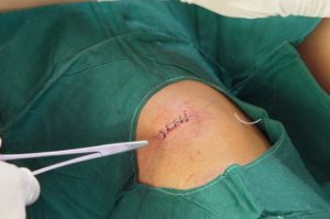
Figure 1. Wound suture
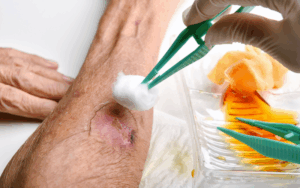
Figure 2. Wound dressing
Technique
Closure by secondary intention effectively addresses wounds on concave head and neck areas particularly those at infection risk.
Results are aesthetic and functional to avoid complex flap or skin graft procedures.
The final scar is less visible in older, lighter-skinned patients, and suitable with other techniques.
Deep sutures eliminate dead space, relieve tension, ensure wound edge alignment, and contribute to final eversion of the wound.
For head and neck wound closure, use 5-0 or 6-0 nonabsorbable Prolene, nylon, or absorbable catgut sutures carefully.
Eversion of skin edges prevents scar depression and place simple suture knots away from wound edges.
Running Locking Suture: Continuous suture with loops locked for added strength.
Vertical Mattress Sutures: It provides strong eversion of edges to prevent depressed scars.
Horizontal Mattress Sutures: It distributes tension over a wider area.
Approximate wound edges to initiate healing, avoid tight sutures that compromise blood flow to cause tissue necrosis, scarring, and unsatisfactory cosmetic outcomes.
Sutures tied tightly today will become tighter tomorrow. Despite careful tissue handling to cause swelling and increased tension at the suture site.
Subcuticular suture procedure:
Sutures can be intradermal either simple or running with the needle placed horizontally in the dermis without skin penetration.
The buried knot in a simple suture minimizes wound tension, while continuous subcuticular stitches allow suture ends to be taped without knots.
Wound closure techniques have advanced to include synthetic sutures, staples, tapes, and adhesives.
Synthetic sutures and standardized materials enhance aesthetic results in engineering.
Topical adhesives, surgical staples, and tapes supplement sutures in wound closure techniques.
Proper materials and technique ensure optimal healing. Four wound healing phases studied at cellular and molecular levels.
The four phases are: Hemostasis, Inflammation, Proliferation, Maturation phase
Initial injury disrupts blood vessels and exposed collagen triggers hemostasis with normal thrombotic function.
Epithelial cell migration occurs in 12-24 hours, with new tissue formation over 10-14 days.
Traumatic wounds
Surgical wounds
Chronic wounds
Cosmetic and Reconstructive Needs
Orthopedic Applications
Burns and Extensive Skin Loss
Ophthalmologic and Otolaryngologic Applications
Infection
Chronic wounds
Delayed presentation
High risk of compartment syndrome
Insufficient debridement
Patient-related factors
Tissue loss in avulsion injuries may delay wound closure for controlling necrotic tissue and debris.
Delayed closure of human and some animal bites may prevent infections.
Restoring skin integrity involves forming a strong scar, proper closure techniques, and sealing to prevent contamination.
Well-aligned wound edges and proper closure methods minimize scarring, restore functionality, and reduce pain and anxiety.
Wounds may partially reopen due to improper technique, tension, or infection, or excessive scar tissue in susceptible individuals.
Poor closure techniques and excessive tension impair blood flow in patients with diabetes, malnutrition, or immune suppression.
Wound Irrigation
Debridement Tools
Clamps
Suturing and Closure
Dressings
Tape or Bandages
Adhesives
Patient Preparation:
Inform the patient about surgery risks, benefits, recovery, pain management, restrictions, and physical therapy.
Ensure understanding before obtaining written informed consent.
A mixture of one part sodium bicarbonate and ten parts local anesthesia with epinephrine can reduce burning during lidocaine infiltration.
Patient Positioning:
Position patient for best wound access and visibility.
Figure 1. Wound suture
Figure 2. Wound dressing
Closure by secondary intention effectively addresses wounds on concave head and neck areas particularly those at infection risk.
Results are aesthetic and functional to avoid complex flap or skin graft procedures.
The final scar is less visible in older, lighter-skinned patients, and suitable with other techniques.
Deep sutures eliminate dead space, relieve tension, ensure wound edge alignment, and contribute to final eversion of the wound.
For head and neck wound closure, use 5-0 or 6-0 nonabsorbable Prolene, nylon, or absorbable catgut sutures carefully.
Eversion of skin edges prevents scar depression and place simple suture knots away from wound edges.
Running Locking Suture: Continuous suture with loops locked for added strength.
Vertical Mattress Sutures: It provides strong eversion of edges to prevent depressed scars.
Horizontal Mattress Sutures: It distributes tension over a wider area.
Approximate wound edges to initiate healing, avoid tight sutures that compromise blood flow to cause tissue necrosis, scarring, and unsatisfactory cosmetic outcomes.
Sutures tied tightly today will become tighter tomorrow. Despite careful tissue handling to cause swelling and increased tension at the suture site.
Subcuticular suture procedure:
Sutures can be intradermal either simple or running with the needle placed horizontally in the dermis without skin penetration.
The buried knot in a simple suture minimizes wound tension, while continuous subcuticular stitches allow suture ends to be taped without knots.
Both our subscription plans include Free CME/CPD AMA PRA Category 1 credits.
Digital Certificate PDF
On course completion, you will receive a full-sized presentation quality digital certificate.
medtigo Simulation
A dynamic medical simulation platform designed to train healthcare professionals and students to effectively run code situations through an immersive hands-on experience in a live, interactive 3D environment.
medtigo Points
medtigo points is our unique point redemption system created to award users for interacting on our site. These points can be redeemed for special discounts on the medtigo marketplace as well as towards the membership cost itself.
Community Forum post/reply = 5 points
*Redemption of points can occur only through the medtigo marketplace, courses, or simulation system. Money will not be credited to your bank account. 10 points = $1.
All Your Certificates in One Place
When you have your licenses, certificates and CMEs in one place, it's easier to track your career growth. You can easily share these with hospitals as well, using your medtigo app.