

Free CME credits
Both our subscription plans include Free CME/CPD AMA PRA Category 1 credits.


Chest radiograph of an infant showing bilateral pulmonary hyperinflation in RSV
The respiratory syncytial virus causes a respiratory tract infection transmitted through droplets and is primarily observed in infants and younger children. It hallmarks acute inflammation, increased mucus production, necrosis of the epithelial cells lining the small airways, and bronchospasm. RSV lower respiratory tract infection can manifest radiographically as bronchiolitis, an obstructive lung illness and pneumonitis.

Respiratory syncytial virus under electron microscope. The virion is variable in shape, and size, with an average diameter between 120-300nm
Seasonal variation in RSV incidence exists; temperate regions have a clear winter-spring predominance, whereas equatorial and tropical climates may have less noticeable spikes with more interseasonal sickness. RSV is considered accountable for over 33 million lower respiratory tract illnesses, 3 million hospitalization, and 199,000 juvenile fatalities worldwide.
Premature newborns, patients with prior pulmonary, cardiac, immunological, and neurologic diseases, and the elderly have much-increased morbidity and death. 90% of children are infected within the first two years of life, while older children and adults are reinfected.

Chest radiograph of a healthy child vs a child with bronchiolitis due to RSV
RSV symptoms are similar to common cold symptoms, such as coughing, sneezing, fever, runny nose, and myalgia. The difference is that the runny nose will be substantially mucusy, with large volumes of mucus. Mucus is typically yellow, green, or gray. When the infected child breathes, they make a whistling or wheezing sound and rarely have red eyes.

Role of neutrophils in the respiratory tract viral infection
The virus quickly spreads throughout the respiratory tract after being inoculated into the nasopharyngeal or conjunctival mucosa. Neutrophil-induced mucus production limits access of viral particles to the epithelium but obstructs airflow by plugging by mucus, cellular waste, and DNA.
Phagocytosis of viral particles and virus-infected cells limits viral spread. Neutrophil extracellular traps capture and deactivate viral particles and damage healthy cells. When the viral cytotoxicity and the host’s cytotoxic response combine to cause the necrosis of respiratory epithelial cells, the host’s inflammatory immune reaction is triggered, including both humoral and cytotoxic T-cell activation.

The pathological changes in the lungs of infant baboon resemble those in infant humans with RSV infection. (H&E: hematoxylin and eosin stain)
The external appearance of intact, fresh lung blocks recovered from RSV-infected animals on days 1, 3, and 5 following infection is shown in A–C. The pathophysiology of airway mucous congestion. The arrows indicate the state of the lung parenchyma in each figure. Lungs demonstrate incrementally greater vascular congestion and edema over time.
D–F demonstrate interstitial pneumonia with worsening inflammation, epithelial damage, and consolidation over time. The inflammatory infiltrate is more subtle on day 1 and found primarily in a peribronchiolar distribution with multifocal infiltration in the bronchiolar epithelium.
Progression to day 5 reveals severe thickening of the interstitium and alveolar collapse because of the worsening inflammatory infiltration. In G–I, there is progressive bronchiolar epithelial damage with sloughing, leading to partial to complete obstruction of some bronchioles with fragmented epithelium mixed with mononuclear inflammatory cells.

Bronchiolitis in an infant with RSV Infection. Lymphocytes infiltrate the bronchiole, and sloughed necrotic material fills and obstructs the bronchiolar lumen. The beginning of the regeneration of bronchiolar epithelium is evident
RSV is a single-stranded RNA virus in the genus Pneumovirus that belongs to the Paramyxoviridae family. It is transmitted from humans by respiratory droplets.
The incubation period following RSV inoculation ranges from 2 to 8 days, with a typical incubation period of 4-6 days, depending on host characteristics such as the patient’s age and first RSV infection.
RSV could develop into a lower respiratory tract infection with various permutations of the classic bronchiolitis symptoms, including tachypnea, wheezes, delayed expiration, and rhonchorous breath sounds.

Bilateral patchy consolidation in a predominantly perihilar distribution extending to the bases. Left lower lobe and right middle lobe confluent consolidation. (Features of RSV pneumonitis in a 9-month-old infant)
Most RSV infections are treated with supportive care. However, passive prophylactic immunization is available for children at risk, such as infants and premature newborns with a history of pulmonary, cardiac, or neuromuscular conditions. The RSV season requires monthly administration of palivizumab.
American Academy of Pediatrics recommends prophylaxis for children in the first year of life with prematurity less than or equal to 29 weeks gestational age, congenital heart disease, chronic lung disease of prematurity, or neuromuscular disorders. Ribavirin is the only antiviral medication approved against RSV in the United States.

X-ray of a child with RSV showing the typical bilateral perihilar fullness of bronchiolitis
RSV infection-related hospitalization in children typically results in complete recovery. They are released after three to four days. Infants at high risk spend more time in the hospital, require more mechanical breathing, and are more likely to be transferred to the intensive care unit.
There is evidence that severe RSV infection and early RSV hospitalization increase the chance of developing recurrent wheezing, childhood asthma, and allergy sensitization.

December 2, 2022

November 7, 2022
Both our subscription plans include Free CME/CPD AMA PRA Category 1 credits.
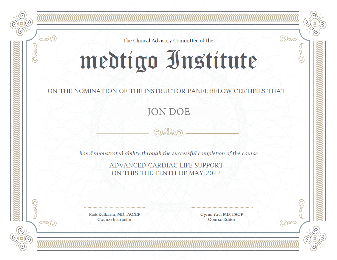
On course completion, you will receive a full-sized presentation quality digital certificate.
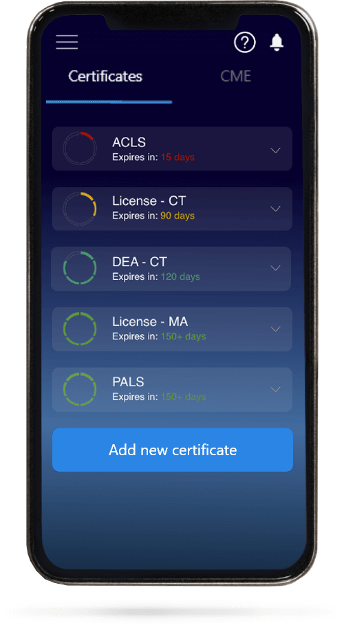
When you have your licenses, certificates and CMEs in one place, it's easier to track your career growth. You can easily share these with hospitals as well, using your medtigo app.


