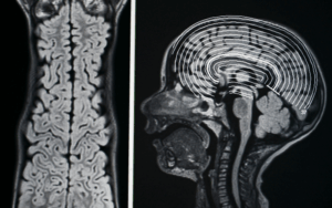
JXG is a uncommon disease from a rare group of non-Langerhans cell histiocytosis that characterize the particular proliferation of certain immune cells in the body in various tissues. In particular, JXG or Therapy-related Myeloid Neoplasms associate with the increased number of histiocytes, the group of immune cells, that cause the development of lesions in the skin and other organs. JXG typically is seen in children during the early years of life, having been developed in the infancy or early childhood stage. It is believed to happen only sometimeswhich makes the precise incidence unknown. Such plaques have several common characteristics, for example, they are usually firm, dome shape and have yellow tinge. Clinical characteristics of the lesions can be diverse- they may be either of small or large sizes while being either solitary or multiple. Skin is usually the most common place that is the affected, but not just the skin, JXG can also involve the eyes, lungs, liver, spleen, and bones. Clinical examination and biopsy are usually used for diagnosis and a histological examination under a macroscopic is performed as a confirmation of the diagnosis as well. The lesion on histopathological examination shows the presence of Touton giant cellswhich are multinucleated cells undergirded by a foamy area of histiocytes.
Epidemiology
More than half of cases are found within the first six months of life with the highest number in the first year. It turned out that Juvenile Xanthogranuloma comprises more of males over females. More reports in males than females are carried out. The disease is classified as rare however as the accurate incidence cases lack clear figures. Skin lesions are usually solitary domed-shaped and the color can be either yellow or red although can be mostly reddish-brown. JXG has the skin as its major target but sometimes it can also involve other organs such as the eyes lungs liver spleen and bones.
Anatomy
Pathophysiology
The primary pathology in JXG is the excessive proliferation of histiocytes. These histiocytes accumulate in various tissues, particularly the skin, leading to the formation of characteristic lesions.
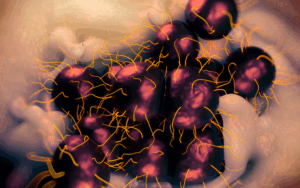
Histopathological examination of JXG lesions often reveals the presence of Touton giant cells.
The Touton cells composed of concentrically nucleated foamy histiocytes constitute a multinucleated giant cell around them. Association with Other Conditions: JXG can occur in many cases although they are clinically linked to neurofibromatosis type 1 (NF1) in some.
Etiology
In the most of Juvenile xanthogranuloma conditions individuals faced the idiopathic state when the disease occurs none- the less having the unclear causal mechanism Although certain links to NF1 or other genetic conditions might be reported in some cases most of the occurrences of this disease happen randomly in those who are not associated with these genetic conditions. Although JXG is rarely considered a hereditary disorder there were some published incidences linking it with various genetic abnormalities. The same disorder causes the inappropriate multiplication of histiocytes in JXG which appear to be a case of immune system dysfunction. In several cases, infectious agents that might induce Juvenile xanthogranuloma and other histiocytic disorders have been considered.
Genetics
Prognostic Factors
The prognosis is connected with the severity of complications including dysfunction of vital organs that in turn are affected by the disease. The most positive prognostic factor for many of cutaneous melanomas is their tendency to regress themselves spare any treatment and this fact still remains unknown. Since early the lesions diagnosed in infants and young children for the very better prognosis and the condition in this case often beneficial is more self-limiting. The treatment of cysts may range from surgical removal to other modalities including topical medicine or more intensive in-clinic therapies depending on the diagnosis and severity of the condition.
Clinical History
Age Group Juvenile xanthogranuloma is the most common among the children below the age of two years, thus it is listed as the diagnosis of the pediatric population most often. The majority of the instances typically occur in children who are less than 2 years old and the peak incidence taking place in the first year of their lives. The word “juvenile”(Juvenile xanthogranuloma) signifies, mostly occurrence at a very young age.
Physical Examination
Age group
Associated comorbidity
It is significant that NF1 is the most common case associated with the narcolepsy. NF1 is a genetic disorder with tumor features arising in nerve cells, skin, and many other parts of the body. In extreme cases JXG has only once been reported in association with hematologic malignancies or hematologic tumors such as leukemia.
Associated activity
Acuity of presentation
JXG in lesions commonly do not produce much pain and are not affected with much discomfort. JXG lesions manifest through slow growth and often remain stable overtime. With prolonged time the size of the lesions may increase and the incident of the new ones still occurs. Lymph nodes affected by JXG results in discoloration and can be yellow to red-brown and usually feel firm.
Differential Diagnoses
Xanthoma
Dermatofibroma
Langerhans Cell Histiocytosis
Granuloma Annulare
Hemangioma
Laboratory Studies
Imaging Studies
Procedures
Histologic Findings
Staging
Treatment Paradigm
Surgical Excision: Resection is sometimes used for cutaneous or benign tumors causing discomfort and cosmetic issues. Corticosteroids: Topical or intralesional corticosteroids may be an option for medium-sized lesions or lesions found in cosmetically-sensitive areas. Ophthalmologic Management: If eye involvement destructs the patients ophthalmologist collaboration becomes a priority for effective treatment plan. Follow-up: The schedule to be followed up regularly to indicate the progress and status of the spots is crucial to determine the treatment requirement.
Avoidance of Trauma: Since JXG lesions can be on the body particularly in children it makes sense not to inflict any harm on the diseased locations of the body which can be avoided while treating. Among other things this also means avoiding any behaviour that could accidentally cause any scratching and rubbing or any movements that can worsen the process of healing. Sun Protection: Sun protection like the wearing of sunscreen and protective clothing is important and should be remembered and especially if the person has got dizzying circles lesions on sun-exposed areas. Differently from the effect on overall skin health it may play the most vital role for the individuals with the wound-healing disturbance. Monitoring and Self-Examination: It is necessary to do skin examination periodically that is searching for morphological changes in the lesion or development of new ones. Skin Care: Soft and sensitive skin care practices including mild soap and moisturizer usages can really benefit the skin health of the person.
Use of Corticosteroids in treating Juvenile Xanthogranuloma
Corticosteroids therapies which are meant to be topically applied and with superficial tissue, the aim is to relieve irritation using the corticosteroids. Injection of glycerine into the lesion area considered in any small local site or which causes the symptoms. Specific corticosteroids are administered in patients with a large exposure and simultaneous extracutaneous indications.
Prednisolone ocular and dexamethasone ocular: Both prednisolone ocular and dexamethasone ocular are class of corticosteroid medications that are mainly applied to various eye diseases where inflammation is a crucial factor. These medications may be prescribed for selected eye symptoms like JXG, and the physician must choose the one that is appropriate.
Biopsy: For a definitive diagnosis with JXG a skin biopsy is usually done. Biopsy suggests microscopic findings. Surgical Excision: Surgical excision sometimes may be done to remove cutaneous lesions that are isolated and which result in cosmetic problems and/or functioning limiting issues.
Diagnosis: A Juvenile Xanthogranulomarequires diagnoses as its initial phase. In this case a complete clinical examination must be performed most often accompanied by the biopsy of the skin-tissue which is commonly carried out for histopathological confirmation. Surgical Excision: Surgical excision of the lesion to the extent possible for the affected skin sites and also those causing irritation symptoms or cosmetic disfigurement. Ongoing Monitoring and Follow-Up: The patient is evaluated thereafter to assess the effectiveness of the given intervention and ongoing monitoring and aftercare is initiated if necessary. Long-Term Management: For affected individuals who did not respond to intervention while disease activity has been in remission the treatment strategy changes to chronic management.
JXG is a uncommon disease from a rare group of non-Langerhans cell histiocytosis that characterize the particular proliferation of certain immune cells in the body in various tissues. In particular, JXG or Therapy-related Myeloid Neoplasms associate with the increased number of histiocytes, the group of immune cells, that cause the development of lesions in the skin and other organs. JXG typically is seen in children during the early years of life, having been developed in the infancy or early childhood stage. It is believed to happen only sometimeswhich makes the precise incidence unknown. Such plaques have several common characteristics, for example, they are usually firm, dome shape and have yellow tinge. Clinical characteristics of the lesions can be diverse- they may be either of small or large sizes while being either solitary or multiple. Skin is usually the most common place that is the affected, but not just the skin, JXG can also involve the eyes, lungs, liver, spleen, and bones. Clinical examination and biopsy are usually used for diagnosis and a histological examination under a macroscopic is performed as a confirmation of the diagnosis as well. The lesion on histopathological examination shows the presence of Touton giant cellswhich are multinucleated cells undergirded by a foamy area of histiocytes.
More than half of cases are found within the first six months of life with the highest number in the first year. It turned out that Juvenile Xanthogranuloma comprises more of males over females. More reports in males than females are carried out. The disease is classified as rare however as the accurate incidence cases lack clear figures. Skin lesions are usually solitary domed-shaped and the color can be either yellow or red although can be mostly reddish-brown. JXG has the skin as its major target but sometimes it can also involve other organs such as the eyes lungs liver spleen and bones.
The primary pathology in JXG is the excessive proliferation of histiocytes. These histiocytes accumulate in various tissues, particularly the skin, leading to the formation of characteristic lesions.
Histopathological examination of JXG lesions often reveals the presence of Touton giant cells.
The Touton cells composed of concentrically nucleated foamy histiocytes constitute a multinucleated giant cell around them. Association with Other Conditions: JXG can occur in many cases although they are clinically linked to neurofibromatosis type 1 (NF1) in some.
In the most of Juvenile xanthogranuloma conditions individuals faced the idiopathic state when the disease occurs none- the less having the unclear causal mechanism Although certain links to NF1 or other genetic conditions might be reported in some cases most of the occurrences of this disease happen randomly in those who are not associated with these genetic conditions. Although JXG is rarely considered a hereditary disorder there were some published incidences linking it with various genetic abnormalities. The same disorder causes the inappropriate multiplication of histiocytes in JXG which appear to be a case of immune system dysfunction. In several cases, infectious agents that might induce Juvenile xanthogranuloma and other histiocytic disorders have been considered.
The prognosis is connected with the severity of complications including dysfunction of vital organs that in turn are affected by the disease. The most positive prognostic factor for many of cutaneous melanomas is their tendency to regress themselves spare any treatment and this fact still remains unknown. Since early the lesions diagnosed in infants and young children for the very better prognosis and the condition in this case often beneficial is more self-limiting. The treatment of cysts may range from surgical removal to other modalities including topical medicine or more intensive in-clinic therapies depending on the diagnosis and severity of the condition.
Age Group Juvenile xanthogranuloma is the most common among the children below the age of two years, thus it is listed as the diagnosis of the pediatric population most often. The majority of the instances typically occur in children who are less than 2 years old and the peak incidence taking place in the first year of their lives. The word “juvenile”(Juvenile xanthogranuloma) signifies, mostly occurrence at a very young age.
It is significant that NF1 is the most common case associated with the narcolepsy. NF1 is a genetic disorder with tumor features arising in nerve cells, skin, and many other parts of the body. In extreme cases JXG has only once been reported in association with hematologic malignancies or hematologic tumors such as leukemia.
JXG in lesions commonly do not produce much pain and are not affected with much discomfort. JXG lesions manifest through slow growth and often remain stable overtime. With prolonged time the size of the lesions may increase and the incident of the new ones still occurs. Lymph nodes affected by JXG results in discoloration and can be yellow to red-brown and usually feel firm.
Xanthoma
Dermatofibroma
Langerhans Cell Histiocytosis
Granuloma Annulare
Hemangioma
Surgical Excision: Resection is sometimes used for cutaneous or benign tumors causing discomfort and cosmetic issues. Corticosteroids: Topical or intralesional corticosteroids may be an option for medium-sized lesions or lesions found in cosmetically-sensitive areas. Ophthalmologic Management: If eye involvement destructs the patients ophthalmologist collaboration becomes a priority for effective treatment plan. Follow-up: The schedule to be followed up regularly to indicate the progress and status of the spots is crucial to determine the treatment requirement.
Ophthalmology
Orthopaedic Surgery
Pediatrics, General
Avoidance of Trauma: Since JXG lesions can be on the body particularly in children it makes sense not to inflict any harm on the diseased locations of the body which can be avoided while treating. Among other things this also means avoiding any behaviour that could accidentally cause any scratching and rubbing or any movements that can worsen the process of healing. Sun Protection: Sun protection like the wearing of sunscreen and protective clothing is important and should be remembered and especially if the person has got dizzying circles lesions on sun-exposed areas. Differently from the effect on overall skin health it may play the most vital role for the individuals with the wound-healing disturbance. Monitoring and Self-Examination: It is necessary to do skin examination periodically that is searching for morphological changes in the lesion or development of new ones. Skin Care: Soft and sensitive skin care practices including mild soap and moisturizer usages can really benefit the skin health of the person.
Oncology, Other
Ophthalmology
Corticosteroids therapies which are meant to be topically applied and with superficial tissue, the aim is to relieve irritation using the corticosteroids. Injection of glycerine into the lesion area considered in any small local site or which causes the symptoms. Specific corticosteroids are administered in patients with a large exposure and simultaneous extracutaneous indications.
Prednisolone ocular and dexamethasone ocular: Both prednisolone ocular and dexamethasone ocular are class of corticosteroid medications that are mainly applied to various eye diseases where inflammation is a crucial factor. These medications may be prescribed for selected eye symptoms like JXG, and the physician must choose the one that is appropriate.
Ophthalmology
Pediatrics, General
Surgery, General
Biopsy: For a definitive diagnosis with JXG a skin biopsy is usually done. Biopsy suggests microscopic findings. Surgical Excision: Surgical excision sometimes may be done to remove cutaneous lesions that are isolated and which result in cosmetic problems and/or functioning limiting issues.
Ophthalmology
Diagnosis: A Juvenile Xanthogranulomarequires diagnoses as its initial phase. In this case a complete clinical examination must be performed most often accompanied by the biopsy of the skin-tissue which is commonly carried out for histopathological confirmation. Surgical Excision: Surgical excision of the lesion to the extent possible for the affected skin sites and also those causing irritation symptoms or cosmetic disfigurement. Ongoing Monitoring and Follow-Up: The patient is evaluated thereafter to assess the effectiveness of the given intervention and ongoing monitoring and aftercare is initiated if necessary. Long-Term Management: For affected individuals who did not respond to intervention while disease activity has been in remission the treatment strategy changes to chronic management.
Both our subscription plans include Free CME/CPD AMA PRA Category 1 credits.
Digital Certificate PDF
On course completion, you will receive a full-sized presentation quality digital certificate.
medtigo Simulation
A dynamic medical simulation platform designed to train healthcare professionals and students to effectively run code situations through an immersive hands-on experience in a live, interactive 3D environment.
medtigo Points
medtigo points is our unique point redemption system created to award users for interacting on our site. These points can be redeemed for special discounts on the medtigo marketplace as well as towards the membership cost itself.
Community Forum post/reply = 5 points
*Redemption of points can occur only through the medtigo marketplace, courses, or simulation system. Money will not be credited to your bank account. 10 points = $1.
All Your Certificates in One Place
When you have your licenses, certificates and CMEs in one place, it's easier to track your career growth. You can easily share these with hospitals as well, using your medtigo app.