ECFMG Announces 2026 Certification Pathways, New Application System, and Fee Updates for International Medical Graduates
March 27, 2026


Background
Scrofula, historically referred to as the “King’s Evil,” is a term used to describe a specific manifestation of tuberculosis (TB) affecting the lymph nodes in the neck. It primarily involves the cervical lymph nodes and is characterized by the development of swollen, firm, and sometimes painful masses in the neck.
These masses, known as scrofulous nodes or tuberculous lymphadenitis, are a result of the immune system’s response to Mycobacterium tuberculosis, the bacteria responsible for tuberculosis.
Scrofula gained significant attention throughout history due to its association with monarchy and nobility. It was believed that the touch of a monarch had the power to heal scrofulous individuals, leading to the term “King’s Evil.” Monarchs, particularly in Europe, were known to perform the “royal touch” ceremony as a ritualistic form of healing for those afflicted with scrofula.
Epidemiology
The epidemiology of scrofula, also known as tuberculous lymphadenitis, has evolved over time due to advancements in medical knowledge, improved hygiene practices, and the availability of effective tuberculosis (TB) treatments. Scrofula is a manifestation of extrapulmonary tuberculosis, which refers to TB infections occurring outside of the lungs.
Here are some key points regarding the epidemiology of scrofula:
Anatomy
Pathophysiology
Scrofula, also known as tuberculous lymphadenitis, is a manifestation of extrapulmonary tuberculosis (TB) that primarily affects the lymph nodes in the neck. The pathophysiology of scrofula involves the spread of Mycobacterium tuberculosis, the bacterium responsible for TB, through the lymphatic system. Here’s an overview of the pathophysiological processes involved in scrofula:
Inhalation of Tuberculosis Bacteria: The pathophysiological process begins when a person inhales airborne droplets containing M. tuberculosis. These droplets are released into the air when an infected individual with pulmonary TB coughs, sneezes, or talks.
Lung Infection: Once inhaled, M. tuberculosis can establish an infection in the lungs, leading to primary pulmonary tuberculosis. The immune system responds to the infection by forming granulomas, which are organized collections of immune cells.
Lymphatic Spread: In some cases, the bacteria within the granulomas can spread to the lymphatic system, leading to lymphatic dissemination. The lymphatic vessels serve as a pathway for the bacteria to move from the lungs to other parts of the body.
Lymph Node Involvement: The bacteria can reach the regional lymph nodes in the neck, causing an immune response in these nodes. The bacteria are carried by immune cells within the lymphatic fluid.
Granuloma Formation: Similar to the response in the lungs, the immune system attempts to contain the infection by forming granulomas in the affected lymph nodes. These granulomas consist of immune cells, such as macrophages and T cells, which attempt to control and encapsulate the bacteria.
Enlarged Lymph Nodes: As the immune response intensifies, the lymph nodes become swollen and firm. These enlarged lymph nodes, referred to as scrofulous nodes, can be felt as masses in the neck.
Necrosis and Abscess Formation: In some cases, the granulomas may undergo necrosis (cell death), leading to the formation of abscesses within the lymph nodes. This can result in the softening of the lymph nodes and the potential for abscess drainage through the skin.
Clinical Symptoms: The swollen and sometimes painful lymph nodes can cause discomfort, pain, and visible masses in the neck. Abscesses might drain through sinuses or fistulae, creating external openings on the skin.
Systemic Spread (Rare): If left untreated, M. tuberculosis can continue to spread through the bloodstream, potentially causing disseminated tuberculosis involving other organs.
Etiology
The etiology of scrofula is rooted in infection with Mycobacterium tuberculosis, the bacterium responsible for tuberculosis (TB). Scrofula, also known as tuberculous lymphadenitis, is a manifestation of extrapulmonary tuberculosis, which refers to TB infections occurring outside of the lungs. Here’s an overview of the etiological factors and processes involved in scrofula:
Transmission of M. tuberculosis: Scrofula is primarily caused by the spread of M. tuberculosis bacteria from an initial site of infection, usually in the lungs, to the lymph nodes in the neck. The bacteria are transmitted through inhalation of airborne droplets containing the bacterium, released into the air when an infected person coughs, sneezes, or talks.
Initial Lung Infection: Upon inhalation, M. tuberculosis bacteria can establish an infection in the lungs, leading to pulmonary tuberculosis. The bacteria multiply and trigger an immune response, resulting in the formation of granulomas, which are organized collections of immune cells.
Lymphatic Spread: In some cases, the bacteria within the lung granulomas can spread through the lymphatic vessels, leading to lymphatic dissemination. The lymphatic system is a network of vessels that transport lymph fluid, which contains immune cells and other components, throughout the body.
Lymph Node Infection: As the bacteria reach the regional lymph nodes in the neck through the lymphatic vessels, they trigger an immune response in these nodes. This response leads to the formation of granulomas within the lymph nodes.
Granuloma Formation: The immune system attempts to contain the infection by forming granulomas in the affected lymph nodes. Granulomas are structures composed of immune cells, such as macrophages and T cells, that attempt to encapsulate the bacteria.
Lymph Node Enlargement: The formation of granulomas and the influx of immune cells cause the affected lymph nodes to enlarge and become palpable as firm masses in the neck. These enlarged lymph nodes are characteristic of scrofula.
Necrosis and Abscess Formation: In some cases, the granulomas within the lymph nodes may undergo necrosis (cell death), leading to the formation of abscesses within the nodes. This can result in the softening of the lymph nodes and, in certain instances, abscess drainage through the skin.
The etiology of scrofula is directly linked to the pathogen M. tuberculosis and its ability to spread through the lymphatic system, resulting in the characteristic enlargement of lymph nodes in the neck. Early diagnosis, appropriate treatment with anti-tuberculosis medications, and adherence to treatment protocols are essential for managing scrofula and preventing the progression of the disease.
Genetics
Prognostic Factors
The prognosis of scrofula, also known as tuberculous lymphadenitis, is generally favorable with appropriate and timely medical intervention. With the advent of effective tuberculosis (TB) treatment, scrofula can be successfully managed, and most individuals can achieve complete recovery. However, the prognosis can be influenced by several factors:
Timely Diagnosis and Treatment: Early diagnosis and prompt initiation of anti-TB treatment play a crucial role in achieving a favorable outcome. Delayed treatment can lead to complications and potential spread of the infection.
Drug Sensitivity and Resistance: The response to treatment depends on the sensitivity of the TB bacteria to the prescribed medications. Drug-resistant TB strains can complicate treatment and may require alternative drug regimens.
Adherence to Treatment: Adherence to the full course of anti-TB medications is essential to ensure successful treatment and prevent recurrence. Incomplete treatment can lead to drug resistance and treatment failure.
Abscess Formation and Complications: Scrofula cases with abscess formation might require drainage and proper wound care. Complications related to abscesses, such as secondary infections, can impact the prognosis.
Underlying Health Conditions: The patient’s overall health and immune system function can affect their ability to fight off the infection and respond to treatment.
Immunocompromised Individuals: Individuals with weakened immune systems, such as those with HIV/AIDS, may have a more complex course of treatment and a potentially higher risk of complications.
Treatment Adverse Effects: Anti-TB medications can sometimes cause side effects, which might affect the patient’s adherence to treatment and overall well-being.
Follow-Up and Monitoring: Regular follow-up appointments with healthcare providers are crucial to monitor treatment progress, assess response to therapy, and address any concerns or complications.
Patient Adherence and Lifestyle: Patients who adhere to treatment recommendations, maintain a healthy lifestyle, and follow preventive measures can contribute to a better prognosis.
Public Health Factors: The prevalence of TB in the community, access to healthcare, and public health interventions can impact the overall prognosis.
Clinical History
Clinical history
The clinical history of a patient with suspected scrofula provides valuable information that helps healthcare professionals understand the patient’s symptoms, medical background, and potential risk factors. Gathering a comprehensive clinical history involves asking relevant questions to uncover details about the onset, progression, and associated factors of the condition. Here are key components of the clinical history for a patient with suspected scrofula:
Physical Examination
Physical examination
During a physical examination for suspected scrofula, healthcare professionals assess the patient’s neck and lymph nodes for signs of swelling, tenderness, and other characteristic features. Here’s what a physical examination for scrofula might involve:
General Observation: Observe the patient’s overall appearance, posture, and any visible discomfort.
Neck Inspection: Inspect the neck for any visible swelling, asymmetry, or protruding masses.
Note the presence of any redness, warmth, or skin changes overlying the affected lymph nodes.
Palpation of Lymph Nodes: Gently palpate the lymph nodes in the neck. Scrofulous nodes are typically firm, enlarged, and sometimes tender.
Check for the number, size, consistency, and mobility of the lymph nodes.
Location of Enlargement: Note the specific locations of the enlarged lymph nodes. Scrofulous nodes are often found in the cervical (neck) region.
Tenderness: Assess for tenderness or pain upon palpation of the lymph nodes. Tender nodes can indicate an inflammatory or infectious process.
Skin Changes: Examine the skin overlying the lymph nodes for any signs of inflammation, abscess formation, or draining sinuses.
Mobility: Evaluate the mobility of the lymph nodes. Scrofulous nodes may be relatively mobile within the surrounding tissues.
Systemic Examination: Perform a thorough examination of other body systems to assess for signs of systemic illness or associated conditions.
Assessment of Associated Symptoms: Inquire about any symptoms that might be associated with scrofula, such as fever, night sweats, weight loss, or respiratory symptoms.
Medical History: Review the patient’s medical history, including any history of TB exposure, previous TB infection, or other relevant medical conditions.
Comparison with Unaffected Side: Compare the affected side of the neck with the unaffected side to assess for asymmetry and differences in lymph node size or tenderness.
Documentation: Document findings regarding the size, location, tenderness, and consistency of the enlarged lymph nodes.
Age group
Associated comorbidity
Associated activity
Acuity of presentation
Differential Diagnoses
The differential diagnosis of scrofula involves considering other conditions that can cause swollen lymph nodes in the neck, as well as different manifestations of tuberculosis (TB) and other infections. Proper differentiation is crucial to ensure accurate diagnosis and appropriate treatment. Here are some conditions that might be included in the differential diagnosis of scrofula:
Lymphadenitis of Other Etiologies:
Bacterial Lymphadenitis: Infections caused by various bacteria, such as Staphylococcus aureus or Streptococcus pyogenes, can lead to swollen lymph nodes.
Viral Lymphadenitis: Viruses like Epstein-Barr virus (EBV), cytomegalovirus (CMV), and HIV can cause lymph node enlargement.
Cat-Scratch Disease:
A bacterial infection caused by Bartonella henselae, typically contracted through cat scratches or bites, leading to swollen and painful lymph nodes, especially in the neck.
Lymphoma:
Lymphomas are cancers that originate in lymphocytes. Non-Hodgkin lymphoma and Hodgkin lymphoma can present with painless lymph node enlargement.
Salivary Gland Infections:
Infections of the salivary glands, such as mumps, can cause swollen glands in the neck area.
Sarcoidosis:
A systemic disease involving the growth of granulomas in various organs, including the lymph nodes.
Tuberculous Lymphadenitis (Scrofula):
While scrofula itself is a form of tuberculous lymphadenitis, other forms of extrapulmonary TB, such as pleural TB or skeletal TB, might also cause lymph node involvement.
Nontuberculous Mycobacterial Infections:
Infections caused by mycobacterial species other than Mycobacterium tuberculosis can also lead to lymphadenitis, known as nontuberculous mycobacterial lymphadenitis.
Lymph Node Metastasis:
Enlarged lymph nodes might be due to metastatic spread of cancer from nearby or distant primary sites.
Kikuchi-Fujimoto Disease:
A rare, self-limited condition characterized by swollen lymph nodes, fever, and fatigue. The cause is unclear, but it might have an immune-mediated basis.
Connective Tissue Disorders:
Conditions like systemic lupus erythematosus (SLE) or rheumatoid arthritis (RA) can sometimes cause lymphadenopathy.
Toxoplasmosis:
An infection caused by the parasite Toxoplasma gondii, which can lead to lymph node enlargement.
Inflammatory Conditions:
Chronic inflammatory conditions like sarcoidosis or autoimmune disorders can cause lymphadenopathy.
Accurate diagnosis often involves a combination of clinical evaluation, medical history, imaging studies, and sometimes laboratory tests (such as lymph node biopsy) to identify the underlying cause of lymph node enlargement. Collaboration between healthcare providers and specialists may be necessary to reach a definitive diagnosis and initiate appropriate treatment.
Laboratory Studies
Imaging Studies
Procedures
Histologic Findings
Staging
Treatment Paradigm
The management of scrofula, also known as tuberculous lymphadenitis, involves a combination of medical interventions to treat the underlying tuberculosis (TB) infection and address the associated lymph node inflammation and swelling. The goal of treatment is to effectively eliminate the TB bacteria, reduce symptoms, and prevent complications. Here’s an overview of the management approach for scrofula:
Diagnostic Evaluation: Confirm the diagnosis of scrofula through clinical examination, medical history, imaging studies, and potentially laboratory tests (such as lymph node biopsy or culture) to identify the presence of Mycobacterium tuberculosis.
Anti-Tuberculosis Medications: Initiate treatment with a combination of anti-TB medications. The standard regimen typically includes a combination of isoniazid, rifampin, pyrazinamide, and ethambutol for an initial phase of treatment, followed by a continuation phase with isoniazid and rifampin.
The choice of medications and treatment duration may vary based on the patient’s age, overall health, drug sensitivity testing, and local treatment guidelines.
Directly Observed Therapy (DOT): Consider implementing directly observed therapy (DOT) to ensure patient adherence to the medication regimen. In DOT, a healthcare provider or a trained observer watches the patient take each dose of medication.
Follow-Up Monitoring: Regularly monitor the patient’s response to treatment through clinical evaluation, imaging studies, and laboratory tests to assess the reduction in lymph node size, resolution of symptoms, and treatment effectiveness.
Addressing Abscesses: If abscesses or draining sinuses are present, they might require drainage, which can be performed by a healthcare professional. Proper wound care and hygiene are essential to prevent secondary infections.
Pain Management: If the patient experiences pain or discomfort, pain management with over-the-counter or prescription pain relievers may be recommended.
Nutritional Support: Ensure that the patient receives proper nutrition to support their immune system and overall health.
Adherence Support: Provide education to the patient about the importance of completing the entire course of anti-TB medications to prevent drug resistance and recurrence of infection.
Contact Tracing: Identify and screen close contacts of the patient to determine if others have been exposed to TB. Those found to be infected may require treatment as well.
Prevention Measures: Promote preventive measures to reduce the risk of TB transmission, including proper respiratory hygiene, ventilation of living spaces, and awareness of TB symptoms.
Immunization: Depending on the patient’s age and region, consider administering the Bacillus Calmette-Guérin (BCG) vaccine, which can provide partial protection against TB.
Multidisciplinary Care: Collaboration with infectious disease specialists, pulmonologists, and other healthcare providers can ensure comprehensive care and treatment.
by Stage
by Modality
Chemotherapy
Radiation Therapy
Surgical Interventions
Hormone Therapy
Immunotherapy
Hyperthermia
Photodynamic Therapy
Stem Cell Transplant
Targeted Therapy
Palliative Care
Medication
Future Trends
References
https://www.ncbi.nlm.nih.gov/books/NBK557700/
Scrofula, historically referred to as the “King’s Evil,” is a term used to describe a specific manifestation of tuberculosis (TB) affecting the lymph nodes in the neck. It primarily involves the cervical lymph nodes and is characterized by the development of swollen, firm, and sometimes painful masses in the neck.
These masses, known as scrofulous nodes or tuberculous lymphadenitis, are a result of the immune system’s response to Mycobacterium tuberculosis, the bacteria responsible for tuberculosis.
Scrofula gained significant attention throughout history due to its association with monarchy and nobility. It was believed that the touch of a monarch had the power to heal scrofulous individuals, leading to the term “King’s Evil.” Monarchs, particularly in Europe, were known to perform the “royal touch” ceremony as a ritualistic form of healing for those afflicted with scrofula.
The epidemiology of scrofula, also known as tuberculous lymphadenitis, has evolved over time due to advancements in medical knowledge, improved hygiene practices, and the availability of effective tuberculosis (TB) treatments. Scrofula is a manifestation of extrapulmonary tuberculosis, which refers to TB infections occurring outside of the lungs.
Here are some key points regarding the epidemiology of scrofula:
Scrofula, also known as tuberculous lymphadenitis, is a manifestation of extrapulmonary tuberculosis (TB) that primarily affects the lymph nodes in the neck. The pathophysiology of scrofula involves the spread of Mycobacterium tuberculosis, the bacterium responsible for TB, through the lymphatic system. Here’s an overview of the pathophysiological processes involved in scrofula:
Inhalation of Tuberculosis Bacteria: The pathophysiological process begins when a person inhales airborne droplets containing M. tuberculosis. These droplets are released into the air when an infected individual with pulmonary TB coughs, sneezes, or talks.
Lung Infection: Once inhaled, M. tuberculosis can establish an infection in the lungs, leading to primary pulmonary tuberculosis. The immune system responds to the infection by forming granulomas, which are organized collections of immune cells.
Lymphatic Spread: In some cases, the bacteria within the granulomas can spread to the lymphatic system, leading to lymphatic dissemination. The lymphatic vessels serve as a pathway for the bacteria to move from the lungs to other parts of the body.
Lymph Node Involvement: The bacteria can reach the regional lymph nodes in the neck, causing an immune response in these nodes. The bacteria are carried by immune cells within the lymphatic fluid.
Granuloma Formation: Similar to the response in the lungs, the immune system attempts to contain the infection by forming granulomas in the affected lymph nodes. These granulomas consist of immune cells, such as macrophages and T cells, which attempt to control and encapsulate the bacteria.
Enlarged Lymph Nodes: As the immune response intensifies, the lymph nodes become swollen and firm. These enlarged lymph nodes, referred to as scrofulous nodes, can be felt as masses in the neck.
Necrosis and Abscess Formation: In some cases, the granulomas may undergo necrosis (cell death), leading to the formation of abscesses within the lymph nodes. This can result in the softening of the lymph nodes and the potential for abscess drainage through the skin.
Clinical Symptoms: The swollen and sometimes painful lymph nodes can cause discomfort, pain, and visible masses in the neck. Abscesses might drain through sinuses or fistulae, creating external openings on the skin.
Systemic Spread (Rare): If left untreated, M. tuberculosis can continue to spread through the bloodstream, potentially causing disseminated tuberculosis involving other organs.
The etiology of scrofula is rooted in infection with Mycobacterium tuberculosis, the bacterium responsible for tuberculosis (TB). Scrofula, also known as tuberculous lymphadenitis, is a manifestation of extrapulmonary tuberculosis, which refers to TB infections occurring outside of the lungs. Here’s an overview of the etiological factors and processes involved in scrofula:
Transmission of M. tuberculosis: Scrofula is primarily caused by the spread of M. tuberculosis bacteria from an initial site of infection, usually in the lungs, to the lymph nodes in the neck. The bacteria are transmitted through inhalation of airborne droplets containing the bacterium, released into the air when an infected person coughs, sneezes, or talks.
Initial Lung Infection: Upon inhalation, M. tuberculosis bacteria can establish an infection in the lungs, leading to pulmonary tuberculosis. The bacteria multiply and trigger an immune response, resulting in the formation of granulomas, which are organized collections of immune cells.
Lymphatic Spread: In some cases, the bacteria within the lung granulomas can spread through the lymphatic vessels, leading to lymphatic dissemination. The lymphatic system is a network of vessels that transport lymph fluid, which contains immune cells and other components, throughout the body.
Lymph Node Infection: As the bacteria reach the regional lymph nodes in the neck through the lymphatic vessels, they trigger an immune response in these nodes. This response leads to the formation of granulomas within the lymph nodes.
Granuloma Formation: The immune system attempts to contain the infection by forming granulomas in the affected lymph nodes. Granulomas are structures composed of immune cells, such as macrophages and T cells, that attempt to encapsulate the bacteria.
Lymph Node Enlargement: The formation of granulomas and the influx of immune cells cause the affected lymph nodes to enlarge and become palpable as firm masses in the neck. These enlarged lymph nodes are characteristic of scrofula.
Necrosis and Abscess Formation: In some cases, the granulomas within the lymph nodes may undergo necrosis (cell death), leading to the formation of abscesses within the nodes. This can result in the softening of the lymph nodes and, in certain instances, abscess drainage through the skin.
The etiology of scrofula is directly linked to the pathogen M. tuberculosis and its ability to spread through the lymphatic system, resulting in the characteristic enlargement of lymph nodes in the neck. Early diagnosis, appropriate treatment with anti-tuberculosis medications, and adherence to treatment protocols are essential for managing scrofula and preventing the progression of the disease.
The prognosis of scrofula, also known as tuberculous lymphadenitis, is generally favorable with appropriate and timely medical intervention. With the advent of effective tuberculosis (TB) treatment, scrofula can be successfully managed, and most individuals can achieve complete recovery. However, the prognosis can be influenced by several factors:
Timely Diagnosis and Treatment: Early diagnosis and prompt initiation of anti-TB treatment play a crucial role in achieving a favorable outcome. Delayed treatment can lead to complications and potential spread of the infection.
Drug Sensitivity and Resistance: The response to treatment depends on the sensitivity of the TB bacteria to the prescribed medications. Drug-resistant TB strains can complicate treatment and may require alternative drug regimens.
Adherence to Treatment: Adherence to the full course of anti-TB medications is essential to ensure successful treatment and prevent recurrence. Incomplete treatment can lead to drug resistance and treatment failure.
Abscess Formation and Complications: Scrofula cases with abscess formation might require drainage and proper wound care. Complications related to abscesses, such as secondary infections, can impact the prognosis.
Underlying Health Conditions: The patient’s overall health and immune system function can affect their ability to fight off the infection and respond to treatment.
Immunocompromised Individuals: Individuals with weakened immune systems, such as those with HIV/AIDS, may have a more complex course of treatment and a potentially higher risk of complications.
Treatment Adverse Effects: Anti-TB medications can sometimes cause side effects, which might affect the patient’s adherence to treatment and overall well-being.
Follow-Up and Monitoring: Regular follow-up appointments with healthcare providers are crucial to monitor treatment progress, assess response to therapy, and address any concerns or complications.
Patient Adherence and Lifestyle: Patients who adhere to treatment recommendations, maintain a healthy lifestyle, and follow preventive measures can contribute to a better prognosis.
Public Health Factors: The prevalence of TB in the community, access to healthcare, and public health interventions can impact the overall prognosis.
Clinical history
The clinical history of a patient with suspected scrofula provides valuable information that helps healthcare professionals understand the patient’s symptoms, medical background, and potential risk factors. Gathering a comprehensive clinical history involves asking relevant questions to uncover details about the onset, progression, and associated factors of the condition. Here are key components of the clinical history for a patient with suspected scrofula:
Physical examination
During a physical examination for suspected scrofula, healthcare professionals assess the patient’s neck and lymph nodes for signs of swelling, tenderness, and other characteristic features. Here’s what a physical examination for scrofula might involve:
General Observation: Observe the patient’s overall appearance, posture, and any visible discomfort.
Neck Inspection: Inspect the neck for any visible swelling, asymmetry, or protruding masses.
Note the presence of any redness, warmth, or skin changes overlying the affected lymph nodes.
Palpation of Lymph Nodes: Gently palpate the lymph nodes in the neck. Scrofulous nodes are typically firm, enlarged, and sometimes tender.
Check for the number, size, consistency, and mobility of the lymph nodes.
Location of Enlargement: Note the specific locations of the enlarged lymph nodes. Scrofulous nodes are often found in the cervical (neck) region.
Tenderness: Assess for tenderness or pain upon palpation of the lymph nodes. Tender nodes can indicate an inflammatory or infectious process.
Skin Changes: Examine the skin overlying the lymph nodes for any signs of inflammation, abscess formation, or draining sinuses.
Mobility: Evaluate the mobility of the lymph nodes. Scrofulous nodes may be relatively mobile within the surrounding tissues.
Systemic Examination: Perform a thorough examination of other body systems to assess for signs of systemic illness or associated conditions.
Assessment of Associated Symptoms: Inquire about any symptoms that might be associated with scrofula, such as fever, night sweats, weight loss, or respiratory symptoms.
Medical History: Review the patient’s medical history, including any history of TB exposure, previous TB infection, or other relevant medical conditions.
Comparison with Unaffected Side: Compare the affected side of the neck with the unaffected side to assess for asymmetry and differences in lymph node size or tenderness.
Documentation: Document findings regarding the size, location, tenderness, and consistency of the enlarged lymph nodes.
The differential diagnosis of scrofula involves considering other conditions that can cause swollen lymph nodes in the neck, as well as different manifestations of tuberculosis (TB) and other infections. Proper differentiation is crucial to ensure accurate diagnosis and appropriate treatment. Here are some conditions that might be included in the differential diagnosis of scrofula:
Lymphadenitis of Other Etiologies:
Bacterial Lymphadenitis: Infections caused by various bacteria, such as Staphylococcus aureus or Streptococcus pyogenes, can lead to swollen lymph nodes.
Viral Lymphadenitis: Viruses like Epstein-Barr virus (EBV), cytomegalovirus (CMV), and HIV can cause lymph node enlargement.
Cat-Scratch Disease:
A bacterial infection caused by Bartonella henselae, typically contracted through cat scratches or bites, leading to swollen and painful lymph nodes, especially in the neck.
Lymphoma:
Lymphomas are cancers that originate in lymphocytes. Non-Hodgkin lymphoma and Hodgkin lymphoma can present with painless lymph node enlargement.
Salivary Gland Infections:
Infections of the salivary glands, such as mumps, can cause swollen glands in the neck area.
Sarcoidosis:
A systemic disease involving the growth of granulomas in various organs, including the lymph nodes.
Tuberculous Lymphadenitis (Scrofula):
While scrofula itself is a form of tuberculous lymphadenitis, other forms of extrapulmonary TB, such as pleural TB or skeletal TB, might also cause lymph node involvement.
Nontuberculous Mycobacterial Infections:
Infections caused by mycobacterial species other than Mycobacterium tuberculosis can also lead to lymphadenitis, known as nontuberculous mycobacterial lymphadenitis.
Lymph Node Metastasis:
Enlarged lymph nodes might be due to metastatic spread of cancer from nearby or distant primary sites.
Kikuchi-Fujimoto Disease:
A rare, self-limited condition characterized by swollen lymph nodes, fever, and fatigue. The cause is unclear, but it might have an immune-mediated basis.
Connective Tissue Disorders:
Conditions like systemic lupus erythematosus (SLE) or rheumatoid arthritis (RA) can sometimes cause lymphadenopathy.
Toxoplasmosis:
An infection caused by the parasite Toxoplasma gondii, which can lead to lymph node enlargement.
Inflammatory Conditions:
Chronic inflammatory conditions like sarcoidosis or autoimmune disorders can cause lymphadenopathy.
Accurate diagnosis often involves a combination of clinical evaluation, medical history, imaging studies, and sometimes laboratory tests (such as lymph node biopsy) to identify the underlying cause of lymph node enlargement. Collaboration between healthcare providers and specialists may be necessary to reach a definitive diagnosis and initiate appropriate treatment.
The management of scrofula, also known as tuberculous lymphadenitis, involves a combination of medical interventions to treat the underlying tuberculosis (TB) infection and address the associated lymph node inflammation and swelling. The goal of treatment is to effectively eliminate the TB bacteria, reduce symptoms, and prevent complications. Here’s an overview of the management approach for scrofula:
Diagnostic Evaluation: Confirm the diagnosis of scrofula through clinical examination, medical history, imaging studies, and potentially laboratory tests (such as lymph node biopsy or culture) to identify the presence of Mycobacterium tuberculosis.
Anti-Tuberculosis Medications: Initiate treatment with a combination of anti-TB medications. The standard regimen typically includes a combination of isoniazid, rifampin, pyrazinamide, and ethambutol for an initial phase of treatment, followed by a continuation phase with isoniazid and rifampin.
The choice of medications and treatment duration may vary based on the patient’s age, overall health, drug sensitivity testing, and local treatment guidelines.
Directly Observed Therapy (DOT): Consider implementing directly observed therapy (DOT) to ensure patient adherence to the medication regimen. In DOT, a healthcare provider or a trained observer watches the patient take each dose of medication.
Follow-Up Monitoring: Regularly monitor the patient’s response to treatment through clinical evaluation, imaging studies, and laboratory tests to assess the reduction in lymph node size, resolution of symptoms, and treatment effectiveness.
Addressing Abscesses: If abscesses or draining sinuses are present, they might require drainage, which can be performed by a healthcare professional. Proper wound care and hygiene are essential to prevent secondary infections.
Pain Management: If the patient experiences pain or discomfort, pain management with over-the-counter or prescription pain relievers may be recommended.
Nutritional Support: Ensure that the patient receives proper nutrition to support their immune system and overall health.
Adherence Support: Provide education to the patient about the importance of completing the entire course of anti-TB medications to prevent drug resistance and recurrence of infection.
Contact Tracing: Identify and screen close contacts of the patient to determine if others have been exposed to TB. Those found to be infected may require treatment as well.
Prevention Measures: Promote preventive measures to reduce the risk of TB transmission, including proper respiratory hygiene, ventilation of living spaces, and awareness of TB symptoms.
Immunization: Depending on the patient’s age and region, consider administering the Bacillus Calmette-Guérin (BCG) vaccine, which can provide partial protection against TB.
Multidisciplinary Care: Collaboration with infectious disease specialists, pulmonologists, and other healthcare providers can ensure comprehensive care and treatment.
https://www.ncbi.nlm.nih.gov/books/NBK557700/

Both our subscription plans include Free CME/CPD AMA PRA Category 1 credits.

On course completion, you will receive a full-sized presentation quality digital certificate.
A dynamic medical simulation platform designed to train healthcare professionals and students to effectively run code situations through an immersive hands-on experience in a live, interactive 3D environment.
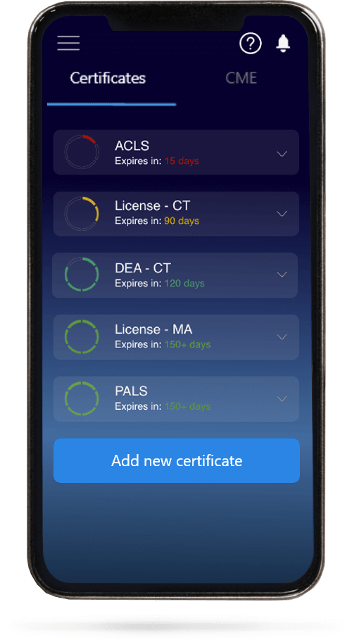
When you have your licenses, certificates and CMEs in one place, it's easier to track your career growth. You can easily share these with hospitals as well, using your medtigo app.