A Milestone Moment: FDA Approves Addyi® for Hypoactive Sexual Desire Disorder in Postmenopausal Women
December 17, 2025


Background
Eagle Syndrome, also known as Eagle’s Syndrome or stylohyoid syndrome, is a rare medical condition characterized by elongation of the styloid process, a slender bony projection located in the neck. This elongation can lead to a range of symptoms due to irritation or compression of nearby structures, including nerves, blood vessels, and the throat.
The condition is named after Dr. Watt W. Eagle, who first described it in the 1930s. The styloid process is a normal anatomical structure in the neck, but in Eagle Syndrome, it becomes abnormally elongated, often exceeding its usual length. This elongated styloid process or the associated calcified ligaments can cause pain and discomfort, particularly when they come into contact with surrounding tissues.
Epidemiology
Eagle Syndrome is considered a relatively rare condition, and its prevalence is not well-established due to limited data and underdiagnosis. However, it’s generally recognized as an uncommon disorder that can affect a small percentage of the population. The exact prevalence can vary among different populations and regions. Eagle Syndrome is more commonly observed in adults, typically in individuals over the age of 30. It appears to be more prevalent in women than in men.
The reasons for these demographic patterns are not entirely clear, but hormonal, anatomical, and developmental factors may play a role. The condition is often misdiagnosed or overlooked because its symptoms, such as throat pain, difficulty swallowing, or referred ear pain, can overlap with those of other more common conditions, including tonsillitis, temporomandibular joint (TMJ) disorders, and ear infections.
Given the rarity of Eagle Syndrome, it’s important for healthcare professionals to be aware of its existence and consider it as a potential diagnosis in individuals with persistent, unexplained throat or neck pain. Accurate diagnosis is crucial for appropriate treatment and management. Because the prevalence and awareness of Eagle Syndrome are still evolving, further research and data collection are needed to better understand its epidemiology, associated risk factors, and optimal treatment approaches.
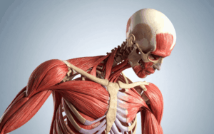
Anatomy
Pathophysiology
The pathophysiology of Eagle Syndrome involves the elongation of the styloid process or calcification of the stylohyoid ligament, both of which are anatomical structures located in the neck. This elongation or calcification can lead to irritation, compression, or impingement on nearby structures, resulting in the characteristic symptoms associated with the condition. Here’s a breakdown of the pathophysiological mechanisms involved in Eagle Syndrome:
Styloid Process Elongation: The styloid process is a slender bony projection that extends down from the temporal bone of the skull, located on each side of the head.
In Eagle Syndrome, the styloid process becomes abnormally elongated, often exceeding its typical length. This elongation can result from various factors, including abnormal bone growth, trauma, or inflammation. The elongated styloid process can come into contact with surrounding structures, such as nerves, blood vessels, and soft tissues.
Stylohyoid Ligament Calcification: The stylohyoid ligament is a thin band of tissue that connects the styloid process to the hyoid bone, a U-shaped bone in the neck.
In some cases of Eagle Syndrome, the stylohyoid ligament becomes calcified or ossified, turning into a rigid structure that can irritate nearby tissues. This calcified ligament can exert pressure on adjacent structures, leading to symptoms like pain and discomfort.
Compression and Irritation: The elongated styloid process or calcified ligament can press against or irritate various nearby structures, including nerves like the glossopharyngeal nerve, vagus nerve, and accessory nerve.
Compression or irritation of these nerves can cause pain, discomfort, and other symptoms that are characteristic of Eagle Syndrome, such as throat pain, difficulty swallowing, referred ear pain, and facial pain.
Referred Pain: The compression and irritation of nerves can lead to referred pain, where discomfort is felt in areas other than the actual site of compression. For example, irritation of the glossopharyngeal nerve can cause pain that radiates to the ear.
Inflammation and Response: The presence of an elongated styloid process or calcified ligament can lead to local inflammation and tissue response. Inflammation can contribute to pain and exacerbate symptoms by sensitizing surrounding tissues. The exact cause of styloid process elongation and stylohyoid ligament calcification in Eagle Syndrome is not fully understood.
Trauma, genetic factors, hormonal influences, and anatomical variations might contribute to these pathophysiological changes. Accurate diagnosis through clinical evaluation, medical history, and imaging studies is important for identifying the underlying cause and tailoring appropriate treatment strategies to relieve symptoms and improve the individual’s quality of life.
Etiology
The etiology of Eagle Syndrome involves the elongation of the styloid process or the calcification of the stylohyoid ligament, both of which are anatomical structures located in the neck. This elongation or calcification can lead to irritation, compression, or impingement on nearby structures, resulting in the characteristic symptoms associated with the condition.
While the exact cause of styloid process elongation or stylohyoid ligament calcification is not fully understood, several factors may contribute to the development of Eagle Syndrome:
It’s important to note that while these factors might contribute to the development of Eagle Syndrome, the condition itself is relatively rare. Additionally, not everyone with elongated styloid processes or calcified ligaments will experience symptoms. The combination of anatomical factors and other influences likely determines whether an individual will develop symptomatic Eagle Syndrome.
Diagnosis is typically based on clinical evaluation, medical history, and imaging studies to visualize the elongated structures and assess their relationship to nearby tissues. Treatment approaches aim to alleviate symptoms and improve the patient’s quality of life, often involving conservative management, medications, and, in severe cases, surgical intervention.
Genetics
Prognostic Factors
The prognosis for individuals with Eagle Syndrome can vary based on several factors, including the severity of their symptoms, the effectiveness of treatment, and individual patient characteristics. While some individuals may experience mild symptoms that respond well to conservative measures, others might have more severe symptoms that require surgical intervention. Here are some key prognostic factors that can influence the outcomes of Eagle Syndrome:
Severity of Symptoms:
The degree of pain, discomfort, and functional impairment caused by Eagle Syndrome can influence the prognosis. Mild symptoms may have a more favorable prognosis, while severe, persistent symptoms might require more intensive treatment.
Duration of Symptoms:
Individuals who seek medical attention and treatment earlier in the course of the condition may have better outcomes. Delayed diagnosis and treatment might lead to chronic pain and complications.
Treatment Approach:
Clinical History
Clinical history
The clinical history of a patient with Eagle Syndrome provides valuable information that aids in the diagnosis and understanding of their condition. Gathering a comprehensive clinical history involves understanding the patient’s symptoms, medical background, and any relevant events that may have led to the onset of their symptoms. Here are the key components of the clinical history for a patient with suspected Eagle Syndrome:
Symptoms: Throat Pain: Inquire about the location, nature, and intensity of the throat pain. Does it occur on one or both sides of the neck?
Difficulty Swallowing: Determine if the patient experiences discomfort or pain while swallowing.
Referred Ear Pain: Ask if they have pain in or around the ear that seems unrelated to any ear issues.
Facial Pain: In some cases, patients may experience facial pain, particularly around the jaw area.
Onset and Duration: When did the symptoms first appear? Is there a specific event or incident that seems to have triggered them?
How long have the symptoms been present? Have they been persistent or intermittent?
Triggers: Ask if there are specific activities, movements, or positions that exacerbate the symptoms, such as swallowing, turning the head, or yawning.
Pain Characteristics: Is the pain sharp, dull, aching, or throbbing?
Does the pain radiate to other areas, such as the ear or jaw?
Medical History: Inquire about any pre-existing medical conditions, surgeries, or trauma that might be relevant to the development of Eagle Syndrome.
Ask about any history of neck injuries, surgeries, or dental procedures, as these can sometimes contribute to the condition.
Physical Examination
Physical examination
A comprehensive physical examination is an important step in the evaluation of a patient with suspected Eagle Syndrome. The examination helps healthcare professionals assess the patient’s symptoms, identify potential signs of the condition, and rule out other possible causes. Here’s what a physical examination for Eagle Syndrome might involve:
Neck Examination:
Palpation: Gently palpate the neck, focusing on the area where the styloid process is located. Note any tenderness or discomfort.
Swelling: Check for any swelling, masses, or abnormal growths in the neck.
Oral and Throat Examination: Observe the oral cavity and throat for any visible abnormalities or signs of inflammation.
Look for any deviation of the uvula (the small, fleshy mass at the back of the throat) from its normal position.
Palpation of Styloid Process: Palpate the styloid process on each side of the head, just beneath the jawline. Note the length and tenderness of the styloid process.
Compare the length of the styloid processes on both sides for any significant asymmetry.
Neck Range of Motion: Ask the patient to move their head and neck in different directions. Observe if certain movements trigger pain or discomfort.
Ear Examination: Examine the ears for signs of infection or inflammation.
Inquire about any ear-related symptoms the patient may have experienced.
Mouth and Jaw Examination: Assess the jaw for any abnormalities, such as pain or clicking sounds during jaw movement.
Observe the patient’s bite and jaw alignment.
Neurological Examination: Assess cranial nerves, particularly the glossopharyngeal nerve (CN IX), vagus nerve (CN X), and accessory nerve (CN XI), which can be affected by Eagle Syndrome.
Imaging Studies: Depending on the clinical findings and the severity of the symptoms, imaging studies such as X-rays, CT scans, or MRI may be ordered to visualize the elongated styloid process or calcified stylohyoid ligament.
Differential Diagnosis: Evaluate the patient for signs that might differentiate Eagle Syndrome from other conditions with similar symptoms.
Associated Symptoms: Assess for other symptoms often associated with Eagle Syndrome, such as referred ear pain, throat discomfort, and difficulty swallowing.
Functional Impact: Inquire about how the symptoms are affecting the patient’s daily life, work, and overall well-being.
Psychosocial Factors: Consider the patient’s emotional well-being, stress levels, and any psychological factors that might contribute to their symptoms.
Age group
Associated comorbidity
Associated activity
Acuity of presentation
Differential Diagnoses
Differential diagnosis
Several conditions can present with symptoms similar to Eagle Syndrome due to overlapping features such as throat pain, difficulty swallowing, and referred ear pain. Accurate diagnosis is important to distinguish Eagle Syndrome from other potential causes of these symptoms. Here are some conditions that might be considered in the differential diagnosis of Eagle Syndrome:
Tonsillitis: Inflammation of the tonsils can cause sore throat, difficulty swallowing, and referred ear pain. Clinical examination and throat cultures can help differentiate tonsillitis from Eagle Syndrome.
Given the wide range of conditions that can produce similar symptoms, a thorough clinical evaluation, medical history review, and potentially imaging studies are crucial to rule out other possible causes and arrive at an accurate diagnosis of Eagle Syndrome. Collaboration among various medical specialists might be necessary to reach a definitive conclusion and recommend appropriate treatment.
Laboratory Studies
Imaging Studies
Procedures
Histologic Findings
Staging
Treatment Paradigm
The treatment of Eagle Syndrome is aimed at relieving symptoms, improving quality of life, and addressing the underlying anatomical issue of elongated styloid processes or calcified stylohyoid ligaments. The treatment approach depends on the severity of symptoms, the impact on the patient’s daily life, and individual patient factors. Here are the main treatment options for Eagle Syndrome:
Conservative Management:
Medications:
Local Injections:
Surgical Interventions:
by Stage
by Modality
Chemotherapy
Radiation Therapy
Surgical Interventions
Hormone Therapy
Immunotherapy
Hyperthermia
Photodynamic Therapy
Stem Cell Transplant
Targeted Therapy
Palliative Care
Medication
Future Trends
References
https://www.ncbi.nlm.nih.gov/books/NBK430789/
Eagle Syndrome, also known as Eagle’s Syndrome or stylohyoid syndrome, is a rare medical condition characterized by elongation of the styloid process, a slender bony projection located in the neck. This elongation can lead to a range of symptoms due to irritation or compression of nearby structures, including nerves, blood vessels, and the throat.
The condition is named after Dr. Watt W. Eagle, who first described it in the 1930s. The styloid process is a normal anatomical structure in the neck, but in Eagle Syndrome, it becomes abnormally elongated, often exceeding its usual length. This elongated styloid process or the associated calcified ligaments can cause pain and discomfort, particularly when they come into contact with surrounding tissues.
Eagle Syndrome is considered a relatively rare condition, and its prevalence is not well-established due to limited data and underdiagnosis. However, it’s generally recognized as an uncommon disorder that can affect a small percentage of the population. The exact prevalence can vary among different populations and regions. Eagle Syndrome is more commonly observed in adults, typically in individuals over the age of 30. It appears to be more prevalent in women than in men.
The reasons for these demographic patterns are not entirely clear, but hormonal, anatomical, and developmental factors may play a role. The condition is often misdiagnosed or overlooked because its symptoms, such as throat pain, difficulty swallowing, or referred ear pain, can overlap with those of other more common conditions, including tonsillitis, temporomandibular joint (TMJ) disorders, and ear infections.
Given the rarity of Eagle Syndrome, it’s important for healthcare professionals to be aware of its existence and consider it as a potential diagnosis in individuals with persistent, unexplained throat or neck pain. Accurate diagnosis is crucial for appropriate treatment and management. Because the prevalence and awareness of Eagle Syndrome are still evolving, further research and data collection are needed to better understand its epidemiology, associated risk factors, and optimal treatment approaches.
The pathophysiology of Eagle Syndrome involves the elongation of the styloid process or calcification of the stylohyoid ligament, both of which are anatomical structures located in the neck. This elongation or calcification can lead to irritation, compression, or impingement on nearby structures, resulting in the characteristic symptoms associated with the condition. Here’s a breakdown of the pathophysiological mechanisms involved in Eagle Syndrome:
Styloid Process Elongation: The styloid process is a slender bony projection that extends down from the temporal bone of the skull, located on each side of the head.
In Eagle Syndrome, the styloid process becomes abnormally elongated, often exceeding its typical length. This elongation can result from various factors, including abnormal bone growth, trauma, or inflammation. The elongated styloid process can come into contact with surrounding structures, such as nerves, blood vessels, and soft tissues.
Stylohyoid Ligament Calcification: The stylohyoid ligament is a thin band of tissue that connects the styloid process to the hyoid bone, a U-shaped bone in the neck.
In some cases of Eagle Syndrome, the stylohyoid ligament becomes calcified or ossified, turning into a rigid structure that can irritate nearby tissues. This calcified ligament can exert pressure on adjacent structures, leading to symptoms like pain and discomfort.
Compression and Irritation: The elongated styloid process or calcified ligament can press against or irritate various nearby structures, including nerves like the glossopharyngeal nerve, vagus nerve, and accessory nerve.
Compression or irritation of these nerves can cause pain, discomfort, and other symptoms that are characteristic of Eagle Syndrome, such as throat pain, difficulty swallowing, referred ear pain, and facial pain.
Referred Pain: The compression and irritation of nerves can lead to referred pain, where discomfort is felt in areas other than the actual site of compression. For example, irritation of the glossopharyngeal nerve can cause pain that radiates to the ear.
Inflammation and Response: The presence of an elongated styloid process or calcified ligament can lead to local inflammation and tissue response. Inflammation can contribute to pain and exacerbate symptoms by sensitizing surrounding tissues. The exact cause of styloid process elongation and stylohyoid ligament calcification in Eagle Syndrome is not fully understood.
Trauma, genetic factors, hormonal influences, and anatomical variations might contribute to these pathophysiological changes. Accurate diagnosis through clinical evaluation, medical history, and imaging studies is important for identifying the underlying cause and tailoring appropriate treatment strategies to relieve symptoms and improve the individual’s quality of life.
The etiology of Eagle Syndrome involves the elongation of the styloid process or the calcification of the stylohyoid ligament, both of which are anatomical structures located in the neck. This elongation or calcification can lead to irritation, compression, or impingement on nearby structures, resulting in the characteristic symptoms associated with the condition.
While the exact cause of styloid process elongation or stylohyoid ligament calcification is not fully understood, several factors may contribute to the development of Eagle Syndrome:
It’s important to note that while these factors might contribute to the development of Eagle Syndrome, the condition itself is relatively rare. Additionally, not everyone with elongated styloid processes or calcified ligaments will experience symptoms. The combination of anatomical factors and other influences likely determines whether an individual will develop symptomatic Eagle Syndrome.
Diagnosis is typically based on clinical evaluation, medical history, and imaging studies to visualize the elongated structures and assess their relationship to nearby tissues. Treatment approaches aim to alleviate symptoms and improve the patient’s quality of life, often involving conservative management, medications, and, in severe cases, surgical intervention.
The prognosis for individuals with Eagle Syndrome can vary based on several factors, including the severity of their symptoms, the effectiveness of treatment, and individual patient characteristics. While some individuals may experience mild symptoms that respond well to conservative measures, others might have more severe symptoms that require surgical intervention. Here are some key prognostic factors that can influence the outcomes of Eagle Syndrome:
Severity of Symptoms:
The degree of pain, discomfort, and functional impairment caused by Eagle Syndrome can influence the prognosis. Mild symptoms may have a more favorable prognosis, while severe, persistent symptoms might require more intensive treatment.
Duration of Symptoms:
Individuals who seek medical attention and treatment earlier in the course of the condition may have better outcomes. Delayed diagnosis and treatment might lead to chronic pain and complications.
Treatment Approach:
Clinical history
The clinical history of a patient with Eagle Syndrome provides valuable information that aids in the diagnosis and understanding of their condition. Gathering a comprehensive clinical history involves understanding the patient’s symptoms, medical background, and any relevant events that may have led to the onset of their symptoms. Here are the key components of the clinical history for a patient with suspected Eagle Syndrome:
Symptoms: Throat Pain: Inquire about the location, nature, and intensity of the throat pain. Does it occur on one or both sides of the neck?
Difficulty Swallowing: Determine if the patient experiences discomfort or pain while swallowing.
Referred Ear Pain: Ask if they have pain in or around the ear that seems unrelated to any ear issues.
Facial Pain: In some cases, patients may experience facial pain, particularly around the jaw area.
Onset and Duration: When did the symptoms first appear? Is there a specific event or incident that seems to have triggered them?
How long have the symptoms been present? Have they been persistent or intermittent?
Triggers: Ask if there are specific activities, movements, or positions that exacerbate the symptoms, such as swallowing, turning the head, or yawning.
Pain Characteristics: Is the pain sharp, dull, aching, or throbbing?
Does the pain radiate to other areas, such as the ear or jaw?
Medical History: Inquire about any pre-existing medical conditions, surgeries, or trauma that might be relevant to the development of Eagle Syndrome.
Ask about any history of neck injuries, surgeries, or dental procedures, as these can sometimes contribute to the condition.
Physical examination
A comprehensive physical examination is an important step in the evaluation of a patient with suspected Eagle Syndrome. The examination helps healthcare professionals assess the patient’s symptoms, identify potential signs of the condition, and rule out other possible causes. Here’s what a physical examination for Eagle Syndrome might involve:
Neck Examination:
Palpation: Gently palpate the neck, focusing on the area where the styloid process is located. Note any tenderness or discomfort.
Swelling: Check for any swelling, masses, or abnormal growths in the neck.
Oral and Throat Examination: Observe the oral cavity and throat for any visible abnormalities or signs of inflammation.
Look for any deviation of the uvula (the small, fleshy mass at the back of the throat) from its normal position.
Palpation of Styloid Process: Palpate the styloid process on each side of the head, just beneath the jawline. Note the length and tenderness of the styloid process.
Compare the length of the styloid processes on both sides for any significant asymmetry.
Neck Range of Motion: Ask the patient to move their head and neck in different directions. Observe if certain movements trigger pain or discomfort.
Ear Examination: Examine the ears for signs of infection or inflammation.
Inquire about any ear-related symptoms the patient may have experienced.
Mouth and Jaw Examination: Assess the jaw for any abnormalities, such as pain or clicking sounds during jaw movement.
Observe the patient’s bite and jaw alignment.
Neurological Examination: Assess cranial nerves, particularly the glossopharyngeal nerve (CN IX), vagus nerve (CN X), and accessory nerve (CN XI), which can be affected by Eagle Syndrome.
Imaging Studies: Depending on the clinical findings and the severity of the symptoms, imaging studies such as X-rays, CT scans, or MRI may be ordered to visualize the elongated styloid process or calcified stylohyoid ligament.
Differential Diagnosis: Evaluate the patient for signs that might differentiate Eagle Syndrome from other conditions with similar symptoms.
Associated Symptoms: Assess for other symptoms often associated with Eagle Syndrome, such as referred ear pain, throat discomfort, and difficulty swallowing.
Functional Impact: Inquire about how the symptoms are affecting the patient’s daily life, work, and overall well-being.
Psychosocial Factors: Consider the patient’s emotional well-being, stress levels, and any psychological factors that might contribute to their symptoms.
Differential diagnosis
Several conditions can present with symptoms similar to Eagle Syndrome due to overlapping features such as throat pain, difficulty swallowing, and referred ear pain. Accurate diagnosis is important to distinguish Eagle Syndrome from other potential causes of these symptoms. Here are some conditions that might be considered in the differential diagnosis of Eagle Syndrome:
Tonsillitis: Inflammation of the tonsils can cause sore throat, difficulty swallowing, and referred ear pain. Clinical examination and throat cultures can help differentiate tonsillitis from Eagle Syndrome.
Given the wide range of conditions that can produce similar symptoms, a thorough clinical evaluation, medical history review, and potentially imaging studies are crucial to rule out other possible causes and arrive at an accurate diagnosis of Eagle Syndrome. Collaboration among various medical specialists might be necessary to reach a definitive conclusion and recommend appropriate treatment.
The treatment of Eagle Syndrome is aimed at relieving symptoms, improving quality of life, and addressing the underlying anatomical issue of elongated styloid processes or calcified stylohyoid ligaments. The treatment approach depends on the severity of symptoms, the impact on the patient’s daily life, and individual patient factors. Here are the main treatment options for Eagle Syndrome:
Conservative Management:
Medications:
Local Injections:
Surgical Interventions:
https://www.ncbi.nlm.nih.gov/books/NBK430789/

Both our subscription plans include Free CME/CPD AMA PRA Category 1 credits.

On course completion, you will receive a full-sized presentation quality digital certificate.
A dynamic medical simulation platform designed to train healthcare professionals and students to effectively run code situations through an immersive hands-on experience in a live, interactive 3D environment.

When you have your licenses, certificates and CMEs in one place, it's easier to track your career growth. You can easily share these with hospitals as well, using your medtigo app.