Entamoeba histolytica is a protozoan parasite known to cause amoebiasis, which most often affects the intestines but may penetrate other tissues, leading to severe consequences.
Epidemiology
E. histolytica is reported worldwide but is more common in developing countries of the tropics and subtropics, where the standard of hygiene is low.
It is believed that each year, the disease affects 50 million individuals, of which 100,000 die; thus, the disease is considered one of the leading killers among parasitic diseases.
Such groups are the travellers, immigrants from endemics areas, institutionalized persons and those who are living in areas with poor hygiene standards.
Anatomy
Pathophysiology
Ingestion: It is contracted when a person takes food or/and drinks containing water that has been contaminated with cysts of E. histolytica.
Excystation: In the small intestine the cysts then develop into the trophozoites, the active invasive form of the parasite.
Colonization: They migrate to the large intestine where they attach themselves to the mucosa usually through specific surface antigens.
Invasion: The E. histolytica trophozoites penetrate the mucosa alter it by the production of enzymes, destroy tissues of the intestinal ulcers and bring inflammation. This can lead to dysentery, mainly associated with diarrhea with blood.
Spread: Sometimes, the trophozoites do not remain within the lumen of the intestine but invade deeper levels, going into the circulatory system. It is observed that the former can subsequently disseminate to other organs; specifically, liver to result in extraintestinal abscesses.
Encystation: Few trophozoites undergo encystation in colon, and thus get passed in the feces, where they can live extracellularly and causes Infection in new host.
Etiology
Entamoeba histolytica is a protozoan parasite which is responsible for the amoebiasis which is an intestinal illness. The etiology consists of the consumption of raw cysts of the parasite through food or water or indirectly through contaminated hands. After entering the host’s intestinal tract, the egg/cysts release the trophozoites which penetrate the intestinal mucosa and create erosions, ulceration and subsequently result into diarrhea, abdominal pains and dysentery. In chronic infections, the parasite migrates to the liver and other organs and forms abscesses.
Genetics
Prognostic Factors
It is tough to predict the outcome of a person infected with Entamoeba histolytica because it depends on several factors such as the severity of the current Infection, and the time at which the diagnosis was made, and when management was commenced.
Clinical History
Age Group:
Children: Entamoeba histolytica affects the children, especially those from unsanitary surroundings, in many ways. The children are more likely to encounter infected foods or water or eat infected foods and their immune system is not well developed as that of the adolescents or adults.
Young Adults (15-35 years): This age group may be at a greater risk for the following reasons: They are more likely to travel especially to endemic areas, and they have behaviours that might contribute to them being exposed to the virus.
Elderly: Those with weak immune systems may be at a higher risk for severe consequences.
Physical Examination
Gastrointestinal tract examination
Liver examination
Respiratory examination
Brain assessment
Cardiac assessment
Age group
Associated comorbidity
Malnutrition: Widespread in endemic areas and intensifies the conditions of amebiasis.
HIV/AIDS: Adjustments for comorbidities included having diabetes, hypertension, chronic steroid use, organ transplant, or immune suppression, the latter being a significant risk factor for severe disease and complications.
Chronic Liver Disease: Patients with predisposing liver diseases have a poorer prognosis if they contract an amoebic liver abscess.
Associated activity
Acuity of presentation
Asymptomatic Carrier State:
Prevalence: Most cases of E. histolytica infections are incidental, and the patient shows no clinical symptoms.
Presentation: Such people become reservoirs of the parasite without any sign of illness. The disease is best diagnosed by observing the cysts in the stool samples.
Intestinal Amebiasis (Amoebic Colitis)
Subacute to Chronic Presentation:
The symptoms appear over time and can be weeks to months in onset.
The clinical signs include abdominal pain, diarrhea and tenesmus which is a feeling of the rectum not being emptied properly.
Including but not limited to diarrhea that is often episodic, may occur with blood or mucus.
Acute Presentation:
Acute severe peritonitis, dysentery, or bloody diarrhea, manifestations of systemic inflammation such as fever.
Some complications arise from severe cases of the illness, and they include toxic megacolon, perforation, and peritonitis.
Amoebic Liver Abscess
Subacute Presentation:
It usually occurs in a longer course of disease, sometimes several weeks after the onset of intestinal Infection.
Some manifestations are abdominal pain, fever, weight loss, and fatigue.
Acute Presentation:
May present with features of acute appendicitis including severe abdominal pain, rigor, high fever and other features of acute systemic Infection.
If ruptured, it causes peritonitis, pleuritis, or some other dangerous conditions,
Extraintestinal Amebiasis
Less Common but Severe:
Parasites can move to other organs and tissues, such as lungs, brain or skin; which are typically fatal in HIV patients.
Infection is localized to an organ although symptoms involve severe pain, fever and other signs of systems involvement from the infected organ.
Differential Diagnoses
Giardia lamblia Infection
Shigella Infection
Salmonella Infection:
Campylobacter Infection
Clostridium difficile Infection
Crohn’s Disease
Ulcerative Colitis
Laboratory Studies
Imaging Studies
Procedures
Histologic Findings
Staging
Treatment Paradigm
Asymptomatic Cysts Carriers: As for those without any symptoms but harbouring these cysts, treatment is advised with a view to avoiding possible spread. The first-line treatment is:
Iodoquinol: Taken orally in a dose of 650 mg three times a day for twenty days.
Paromomycin: Tablet: 25 mg/kg/day orally in divided doses for seven days.
Symptomatic Amoebic Dysentery: Where patients present with clinical features such as dysentery or abdominal pain, for example, the case usually entails:
Metronidazole: 750 mg oral administration thrice daily for 7-10 days.
Alternatively, Tinidazole: 80 mg PO once daily for three days; 2 g PO once daily for three days in severe cases and children.
Following metronidazole or tinidazole therapy, a luminal agent is usually prescribed to ensure complete eradication of the parasite from the intestines:
Iodoquinol: 650 mg oral administration three times a day for 20 days.
Paromomycin: 25 mg /kg/day orally in 3 divided doses for seven days.
Amoebic Liver Abscess: In general, for a liver abscess, patients receive the following:
As initially administered for dysentery, metronidazole or tinidazole.
Once definitive therapy is done, the luminal agent is used as prophylaxis, usually Iodoquinol or Paromomycin.
Alternative Treatments: In some conditions that standard therapies cannot be applied or provide good outcomes Additional chemotherapeutic drugs include Emetine or Dehydroemetines, nevertheless, they are not often used due to side effects.
by Stage
by Modality
Chemotherapy
Radiation Therapy
Surgical Interventions
Hormone Therapy
Immunotherapy
Hyperthermia
Photodynamic Therapy
Stem Cell Transplant
Targeted Therapy
Palliative Care
lifestyle-modifications-in-treating-e-histolytica
Tissue Destruction: E. histolytica has enzymes like proteases, which are lethal to tissues of the host and hence the parasite can penetrate and proliferate in the intestines of the host.
Inflammatory Response: The Infection of the given host initiates an inflammation, which may change the environment of the pathogen’s development. It can transform the tissue nature for the parasite into a better condition and at the same time harm the host tissues.
Microbiome Alteration: Although E. histolytica fails to invade tissues in 50% of affected people, it can change the normal intestine flora by causing dysentery and, thus, affecting the gut microbiome. This can alter nutrient access in the host when immunological responses are triggered.
Acidification: The activity of a parasite at the metabolic level might make localized changes in pH within the host, this has an impact on the organism’s environment and the community of microorganisms.
Role Antibiotics in treating E. histolytica
Metronidazole: This is often the first-line treatment for symptomatic intestinal amoebiasis. It’s effective against the trophozoite form of the parasite.
The recommended adult dose is oral administration of 500-750 mg thrice daily for one week to ten days.
Effectiveness of Luminal agents in treating E. histolytica
Iodoquinol: Another luminal agent which may be employed in the treatment of asymptomatic E.histolytica infection. It is useful in the elimination of the parasites from the intestinal tract.
Paromomycin: Aminoglycoside antibiotic which is used for the treatment of luminal amoebiasis. It is suitable for the non-surgical types of this parasite.
role-of-management-in-treating-e-histolytica
Diagnosis:
Microscopy: Microscopy is done by looking at the stool samples for cysts or trophozoites.
Antigen Detection: For example, in cases where specific antigens are sought, ELISA or any of the immunoassays may be employed.
Molecular Methods: PCR for diagnosing Entamoeba histolytica in pregnant women.
Serology: The serodiagnosis with E. histolytica can be done by detecting antibodies against this parasitic protozoan in the patient’s blood.
Initial Treatment:
Antiparasitic Medications:
Luminal Agents: For example: paromomycin or iodoquinol which can be administered to treat absorptive cases or to eliminate cysts in the intestine.
Systemic Agents: As for example metronidazole or tinidazole, useful in the treatment of both symptomatic parasitic and extraintestinal disease, for example liver abscesses.
Follow-Up and Monitoring:
Symptom Resolution: Monitoring for the resolution of symptoms and any side effects from the medication.
Stool Examination: Having follow-up stool examinations and cysts till they are cleared out.
Imaging: In case of an abscess, repetition of images may be needed after a specific time.
Management of Complications:
Abscesses: Additional treatment for liver abscesses may be with antibiotics, with the occasional requirement of a drainage procedure.
Hydration and Nutritional Support: It is important for patients with severe dysentery to maintain hydration and nutrition.
Prevention and Control:
Hygiene and Sanitation: To avoid Infection; bathing, washing hands thoroughly before eating and consuming safe and clean water.
Education: Results on the measures to take in order not to consume contaminated food and water
Entamoeba histolytica is a protozoan parasite known to cause amoebiasis, which most often affects the intestines but may penetrate other tissues, leading to severe consequences.
E. histolytica is reported worldwide but is more common in developing countries of the tropics and subtropics, where the standard of hygiene is low.
It is believed that each year, the disease affects 50 million individuals, of which 100,000 die; thus, the disease is considered one of the leading killers among parasitic diseases.
Such groups are the travellers, immigrants from endemics areas, institutionalized persons and those who are living in areas with poor hygiene standards.
Ingestion: It is contracted when a person takes food or/and drinks containing water that has been contaminated with cysts of E. histolytica.
Excystation: In the small intestine the cysts then develop into the trophozoites, the active invasive form of the parasite.
Colonization: They migrate to the large intestine where they attach themselves to the mucosa usually through specific surface antigens.
Invasion: The E. histolytica trophozoites penetrate the mucosa alter it by the production of enzymes, destroy tissues of the intestinal ulcers and bring inflammation. This can lead to dysentery, mainly associated with diarrhea with blood.
Spread: Sometimes, the trophozoites do not remain within the lumen of the intestine but invade deeper levels, going into the circulatory system. It is observed that the former can subsequently disseminate to other organs; specifically, liver to result in extraintestinal abscesses.
Encystation: Few trophozoites undergo encystation in colon, and thus get passed in the feces, where they can live extracellularly and causes Infection in new host.
Entamoeba histolytica is a protozoan parasite which is responsible for the amoebiasis which is an intestinal illness. The etiology consists of the consumption of raw cysts of the parasite through food or water or indirectly through contaminated hands. After entering the host’s intestinal tract, the egg/cysts release the trophozoites which penetrate the intestinal mucosa and create erosions, ulceration and subsequently result into diarrhea, abdominal pains and dysentery. In chronic infections, the parasite migrates to the liver and other organs and forms abscesses.
It is tough to predict the outcome of a person infected with Entamoeba histolytica because it depends on several factors such as the severity of the current Infection, and the time at which the diagnosis was made, and when management was commenced.
Age Group:
Children: Entamoeba histolytica affects the children, especially those from unsanitary surroundings, in many ways. The children are more likely to encounter infected foods or water or eat infected foods and their immune system is not well developed as that of the adolescents or adults.
Young Adults (15-35 years): This age group may be at a greater risk for the following reasons: They are more likely to travel especially to endemic areas, and they have behaviours that might contribute to them being exposed to the virus.
Elderly: Those with weak immune systems may be at a higher risk for severe consequences.
Gastrointestinal tract examination
Liver examination
Respiratory examination
Brain assessment
Cardiac assessment
Malnutrition: Widespread in endemic areas and intensifies the conditions of amebiasis.
HIV/AIDS: Adjustments for comorbidities included having diabetes, hypertension, chronic steroid use, organ transplant, or immune suppression, the latter being a significant risk factor for severe disease and complications.
Chronic Liver Disease: Patients with predisposing liver diseases have a poorer prognosis if they contract an amoebic liver abscess.
Asymptomatic Carrier State:
Prevalence: Most cases of E. histolytica infections are incidental, and the patient shows no clinical symptoms.
Presentation: Such people become reservoirs of the parasite without any sign of illness. The disease is best diagnosed by observing the cysts in the stool samples.
Intestinal Amebiasis (Amoebic Colitis)
Subacute to Chronic Presentation:
The symptoms appear over time and can be weeks to months in onset.
The clinical signs include abdominal pain, diarrhea and tenesmus which is a feeling of the rectum not being emptied properly.
Including but not limited to diarrhea that is often episodic, may occur with blood or mucus.
Acute Presentation:
Acute severe peritonitis, dysentery, or bloody diarrhea, manifestations of systemic inflammation such as fever.
Some complications arise from severe cases of the illness, and they include toxic megacolon, perforation, and peritonitis.
Amoebic Liver Abscess
Subacute Presentation:
It usually occurs in a longer course of disease, sometimes several weeks after the onset of intestinal Infection.
Some manifestations are abdominal pain, fever, weight loss, and fatigue.
Acute Presentation:
May present with features of acute appendicitis including severe abdominal pain, rigor, high fever and other features of acute systemic Infection.
If ruptured, it causes peritonitis, pleuritis, or some other dangerous conditions,
Extraintestinal Amebiasis
Less Common but Severe:
Parasites can move to other organs and tissues, such as lungs, brain or skin; which are typically fatal in HIV patients.
Infection is localized to an organ although symptoms involve severe pain, fever and other signs of systems involvement from the infected organ.
Giardia lamblia Infection
Shigella Infection
Salmonella Infection:
Campylobacter Infection
Clostridium difficile Infection
Crohn’s Disease
Ulcerative Colitis
Asymptomatic Cysts Carriers: As for those without any symptoms but harbouring these cysts, treatment is advised with a view to avoiding possible spread. The first-line treatment is:
Iodoquinol: Taken orally in a dose of 650 mg three times a day for twenty days.
Paromomycin: Tablet: 25 mg/kg/day orally in divided doses for seven days.
Symptomatic Amoebic Dysentery: Where patients present with clinical features such as dysentery or abdominal pain, for example, the case usually entails:
Metronidazole: 750 mg oral administration thrice daily for 7-10 days.
Alternatively, Tinidazole: 80 mg PO once daily for three days; 2 g PO once daily for three days in severe cases and children.
Following metronidazole or tinidazole therapy, a luminal agent is usually prescribed to ensure complete eradication of the parasite from the intestines:
Iodoquinol: 650 mg oral administration three times a day for 20 days.
Paromomycin: 25 mg /kg/day orally in 3 divided doses for seven days.
Amoebic Liver Abscess: In general, for a liver abscess, patients receive the following:
As initially administered for dysentery, metronidazole or tinidazole.
Once definitive therapy is done, the luminal agent is used as prophylaxis, usually Iodoquinol or Paromomycin.
Alternative Treatments: In some conditions that standard therapies cannot be applied or provide good outcomes Additional chemotherapeutic drugs include Emetine or Dehydroemetines, nevertheless, they are not often used due to side effects.
Tissue Destruction: E. histolytica has enzymes like proteases, which are lethal to tissues of the host and hence the parasite can penetrate and proliferate in the intestines of the host.
Inflammatory Response: The Infection of the given host initiates an inflammation, which may change the environment of the pathogen’s development. It can transform the tissue nature for the parasite into a better condition and at the same time harm the host tissues.
Microbiome Alteration: Although E. histolytica fails to invade tissues in 50% of affected people, it can change the normal intestine flora by causing dysentery and, thus, affecting the gut microbiome. This can alter nutrient access in the host when immunological responses are triggered.
Acidification: The activity of a parasite at the metabolic level might make localized changes in pH within the host, this has an impact on the organism’s environment and the community of microorganisms.
Metronidazole: This is often the first-line treatment for symptomatic intestinal amoebiasis. It’s effective against the trophozoite form of the parasite.
The recommended adult dose is oral administration of 500-750 mg thrice daily for one week to ten days.
Iodoquinol: Another luminal agent which may be employed in the treatment of asymptomatic E.histolytica infection. It is useful in the elimination of the parasites from the intestinal tract.
Paromomycin: Aminoglycoside antibiotic which is used for the treatment of luminal amoebiasis. It is suitable for the non-surgical types of this parasite.
Diagnosis:
Microscopy: Microscopy is done by looking at the stool samples for cysts or trophozoites.
Antigen Detection: For example, in cases where specific antigens are sought, ELISA or any of the immunoassays may be employed.
Molecular Methods: PCR for diagnosing Entamoeba histolytica in pregnant women.
Serology: The serodiagnosis with E. histolytica can be done by detecting antibodies against this parasitic protozoan in the patient’s blood.
Initial Treatment:
Antiparasitic Medications:
Luminal Agents: For example: paromomycin or iodoquinol which can be administered to treat absorptive cases or to eliminate cysts in the intestine.
Systemic Agents: As for example metronidazole or tinidazole, useful in the treatment of both symptomatic parasitic and extraintestinal disease, for example liver abscesses.
Follow-Up and Monitoring:
Symptom Resolution: Monitoring for the resolution of symptoms and any side effects from the medication.
Stool Examination: Having follow-up stool examinations and cysts till they are cleared out.
Imaging: In case of an abscess, repetition of images may be needed after a specific time.
Management of Complications:
Abscesses: Additional treatment for liver abscesses may be with antibiotics, with the occasional requirement of a drainage procedure.
Hydration and Nutritional Support: It is important for patients with severe dysentery to maintain hydration and nutrition.
Prevention and Control:
Hygiene and Sanitation: To avoid Infection; bathing, washing hands thoroughly before eating and consuming safe and clean water.
Education: Results on the measures to take in order not to consume contaminated food and water
Both our subscription plans include Free CME/CPD AMA PRA Category 1 credits.
Digital Certificate PDF
On course completion, you will receive a full-sized presentation quality digital certificate.
medtigo Simulation
A dynamic medical simulation platform designed to train healthcare professionals and students to effectively run code situations through an immersive hands-on experience in a live, interactive 3D environment.
medtigo Points
medtigo points is our unique point redemption system created to award users for interacting on our site. These points can be redeemed for special discounts on the medtigo marketplace as well as towards the membership cost itself.
Community Forum post/reply = 5 points
*Redemption of points can occur only through the medtigo marketplace, courses, or simulation system. Money will not be credited to your bank account. 10 points = $1.
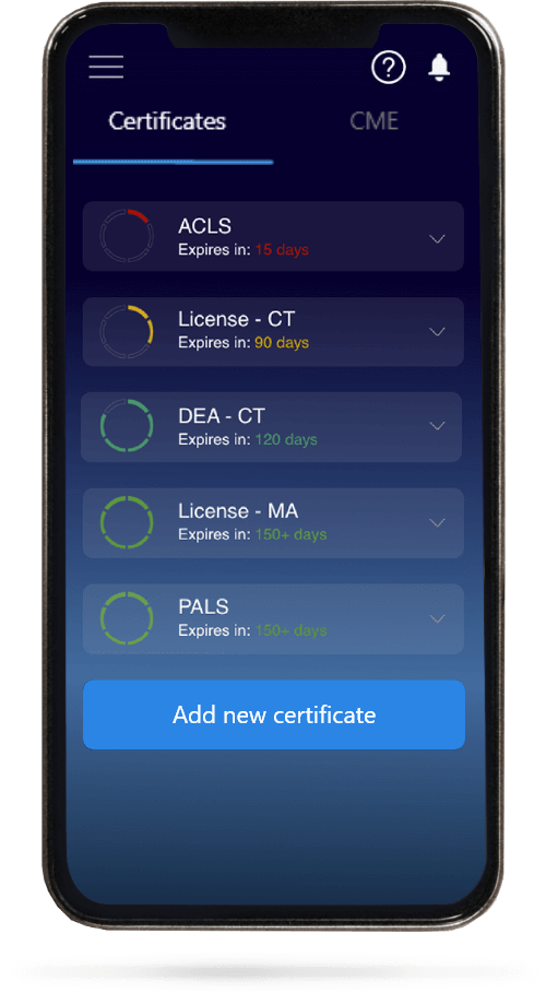
All Your Certificates in One Place
When you have your licenses, certificates and CMEs in one place, it's easier to track your career growth. You can easily share these with hospitals as well, using your medtigo app.