Rising Adoption of Continuous Glucose Monitors in Medicare Advantage: Trends from 2021–2023
April 13, 2026


Background
Peripheral Vascular Disease (PVD) includes peripheral artery disease (PAD) or peripheral arterial disease, characterized by the narrowing or blockage of blood vessels that supply blood to the extremities, usually the legs and feet. This disease is primarily caused by atherosclerosis, a condition where fatty deposits build up on the inner walls of arteries, leading to reduced blood flow to the affected areas.
The primary cause of PVD is atherosclerosis, a progressive condition where cholesterol, fat, and other substances accumulate on the arterial walls, forming plaques. These plaques can restrict blood flow and, in severe cases, lead to complete blockages. Lifestyle changes to manage risk factors, regular medical check-ups, and early intervention are crucial for preventing the progression of PVD.
Epidemiology
The prevalence of PVD increases with age. It is more common in older adults. Estimates suggest that a significant percentage of the population, especially those over 70, may have some degree of peripheral arterial disease. The incidence of PVD tends to rise with age, but it can affect individuals at various life stages. The exact incidence rates can vary based on geographical location, population demographics, and the prevalence of risk factors.
PVD is generally more prevalent in men than in women. However, women are also at risk, especially as they age. The risk of PVD increases substantially with advancing age. Developed countries with higher rates of cardiovascular risk factors may experience a higher prevalence of PVD. African Americans exhibit an odds ratio of 2.12 for peripheral artery disease compared to non-Hispanic whites when adjusting for recognized risk factors.
Anatomy
Pathophysiology
Peripheral vascular disease primarily arises from advancing atherosclerotic disease, leading to dysfunction in both macro and microvascular systems. While peripheral artery disease typically impacts the vascular beds of the lower extremities, larger arteries, such as the abdominal aorta and iliac arteries, are frequently affected. The pathophysiology of atherosclerosis involves a complex inflammatory response encompassing various vascular cells, cholesterol, thrombotic factors, and inflammatory molecules. The initiation of atherosclerosis involves the accumulation of lipoproteins within the intimal layer of large arteries.
This presence of lipoproteins within the endothelium triggers lipid oxidation and a cytokine response, leading to the infiltration of macrophages and lymphocytes. Macrophages consume these oxidized lipids, forming foam cells that contribute to the development of fatty streaks. While not clinically significant initially, these fatty streaks can progress into advanced plaques comprising necrotic lipid cores and smooth muscle cells (SMC). Endothelial cells and SMC release growth factors and cytokines, prompting the migration of SMC to the luminal side of the plaque, facilitating extracellular matrix synthesis, and eventually forming a fibrous plaque.
The stability of fibrous plaques is principally determined by their composition, with more vulnerable plaques having a thinner fibrous cap and a higher number of inflammatory cells. The accumulation of atherosclerotic plaque occurs gradually over decades within the vessel wall. This plaque buildup leads to vascular stenosis and frequent dilation to optimize end-organ perfusion. As the vessel’s dilation capacity reaches its maximum, plaque accumulation continues, further compromising the lumen and occasionally causing critical narrowing of the artery.
With progressive narrowing and obstruction, collateral circulatory beds often develop to maintain tissue viability and distal perfusion. However, these collateral circulatory pathways cannot fully compensate for the blood supply provided by a healthy vessel. Intermittent claudication occurs when blood flow distal to the occlusion is significantly compromised, resulting in fixed oxygen delivery that cannot meet oxygen demand. The most severe manifestation of PAD is critical limb ischemia, defined by limb pain at rest or the impending loss of the limb.
Etiology
Advanced Age
BMI greater than 30
Diabetes Mellitus
HIV
Hypertension
High Cholesterol
Family History of Cardiovascular Disease
Genetics
Prognostic Factors
Clinical History
The clinical manifestation of Peripheral Artery Disease (PAD) is often contingent upon the severity of arterial insufficiency and the coexistence of comorbid conditions, which can modify or obscure the symptoms of underlying vascular disease. Atypical presentations of PAD can arise when patients have pre-existing conditions such as spinal stenosis, lumbosacral disease, or advanced diabetes mellitus, all of which may influence pain perception.
Atypical pain is characterized by sensations not related to physical activity, occurring at rest and during exertion, lasting more than 10 minutes after exercise cessation. Pseudo-claudication is defined as neuropathic pain seen in patients with spinal stenosis and can be distinguished from PAD through a comprehensive physical examination. Patients experiencing pseudo-claudication typically report pain marked by paresthesias and weakness, regardless of the level of physical activity.
This pain is usually alleviated by sitting down or adjusting body positioning rather than requiring rest. The prevalence of asymptomatic PAD may partly be attributed to older individuals misinterpreting their symptoms as normal aging processes. Moreover, patients with mild to moderate PAD may be incapable of exercising to a level that demands significant oxygen supply, resulting in a lack of a mismatch between supply and demand. Consequently, these patients remain asymptomatic.
Physical Examination
The physical examination commences with a comprehensive inspection, focusing on specific indicators such as fingernail tar, suggestive of cigarette smoking, and identifying scars from prior vascular surgeries and the presence of amputations. A targeted cardiovascular examination initiates with a pulse assessment to ascertain rate, rhythm, and strength.
Chest auscultation is then performed to evaluate for pulmonary conditions such as chronic obstructive pulmonary disease and pulmonary fibrosis, along with an assessment for heart sounds or murmurs. A neurological examination is imperative to assess for pseudo-claudication.
Limb examination includes a thorough assessment for pulselessness, muscular atrophy, pallor, cool or cyanotic skin, and pain upon palpation. Lower extremity ulcers may be categorized as arterial, venous, neuropathic, or a combination. Ulcers resulting from arterial insufficiency are typically tender and exhibit ragged borders, a dry base, and pale or necrotic centers.
Age group
Associated comorbidity
Associated activity
Acuity of presentation
Differential Diagnoses
Chronic venous insufficiency
Deep venous thrombosis
Baker cyst
Peripheral neuropathy
Spinal stenosis
Nerve root compression
Laboratory Studies
Imaging Studies
Procedures
Histologic Findings
Staging
Treatment Paradigm
Patients diagnosed with peripheral vascular disease (PAD) necessitate a thoughtful approach that considers factors such as age, risk factors, disease severity, and functional status. The management of PAD is broadly categorized into two objectives: reducing cardiovascular events and alleviating symptoms. Aggressive modification of risk factors is crucial for lowering cardiovascular risk. Quitting smoking, in particular, is instrumental in reducing the progression of PAD, cardiovascular events like myocardial infarction and stroke, and critical limb ischemia.
Educating patients and employing behavioral therapy, nicotine replacement therapy, or pharmacological interventions can effectively reduce smoking and enhance cardiovascular outcomes. Statin therapy has demonstrated efficacy in diminishing cardiovascular events and all-cause mortality while decreasing the need for revascularization. Hence, statins should be routinely incorporated into the treatment plan for patients diagnosed with PAD.
by Stage
by Modality
Chemotherapy
Radiation Therapy
Surgical Interventions
Patients experiencing debilitating symptoms unresponsive to risk factor modification and pharmacological therapy and exercise may be considered for surgical, endovascular, or a combination of both interventions.
The criteria for intervention include individuals with incapacitating claudication that hinders daily activities and those requiring limb salvage due to critical limb ischemia, characterized by ischemic pain at rest, ulceration, and gangrene.
The choice between surgical and percutaneous intervention relies on various factors, such as the patient’s functional status and surgical risk, the proficiency of the operator, the anatomical location and extent of the disease, the presence of multifocal vascular lesions, and the patient’s personal preferences.
Hormone Therapy
Immunotherapy
Hyperthermia
Photodynamic Therapy
Stem Cell Transplant
Targeted Therapy
Palliative Care
Administration with a pharmaceutical agent
Pharmacological management of intermittent claudication (IC) may be considered for patients who have not experienced improvement with exercise therapy and risk factor modification. Two approved medications for treating IC are naftidrofuryl and cilostazol.
Cilostazol, acting as a phosphodiesterase type 3 inhibitor, exhibits antiplatelet effects, vasodilatory properties, and the ability to inhibit smooth muscle cell proliferation. A meta-analysis involving over 2000 patients revealed that individuals using cilostazol experienced significantly longer pain-free and total walking distances.
On the other hand, naftidrofuryl, a 5-hydroxytryptamine-2-receptor antagonist, inhibits glucose uptake and enhances adenosine triphosphate levels. Notably, it has fewer side effects than cilostazol and should be considered when available. Daily aspirin is recommended as part of overall cardiovascular care; however, there must be a consensus on the most effective dosage.
Medication
75
mg
Tablet
Orally
every day
12 - 48
mg
Tablet
Orally
divided into 3 to 4 doses every day
2 gm each day initially
Maintain the dose at 800-1200 mg each day
Keep the maximum dose upto 2 gm in daily divided doses
Indicated for Peripheral vascular disease
120 mcg orally every day in divided three times a day
Primary pulmonary hypertension
Initial dose: 60 mcg orally every day in divided three times a day
It may enhance to 180 mcg orally every day in divided three-four times a day
100 to 200 mg thrice a day
Buflomedil is a vasodilator medication that improves muscle cell metabolism, platelet aggregation inhibition, and the deformability of red blood cells (RBC)
It is a mild calcium channel blocker and an inhibitor of alpha-adrenoceptors
The recommended usual dose is 300-600 mg via oral administration in a day
The recommended usual IM dose is up to max 100 mg in a day
The recommended usual IV dose via slow IV injection is up to max 200 mg in a day or up to max 400 mg in a day by infusion
Dose Adjustments
Limited data is available
Indicated in the treatment of peripheral vascular disease and cerebrovascular insufficiency related mental deterioration
Typically, a divided daily dose of 3–6 mg is advised
Dose Adjustments
Limited data is available
Future Trends
References
Peripheral Vascular Disease (PVD) includes peripheral artery disease (PAD) or peripheral arterial disease, characterized by the narrowing or blockage of blood vessels that supply blood to the extremities, usually the legs and feet. This disease is primarily caused by atherosclerosis, a condition where fatty deposits build up on the inner walls of arteries, leading to reduced blood flow to the affected areas.
The primary cause of PVD is atherosclerosis, a progressive condition where cholesterol, fat, and other substances accumulate on the arterial walls, forming plaques. These plaques can restrict blood flow and, in severe cases, lead to complete blockages. Lifestyle changes to manage risk factors, regular medical check-ups, and early intervention are crucial for preventing the progression of PVD.
The prevalence of PVD increases with age. It is more common in older adults. Estimates suggest that a significant percentage of the population, especially those over 70, may have some degree of peripheral arterial disease. The incidence of PVD tends to rise with age, but it can affect individuals at various life stages. The exact incidence rates can vary based on geographical location, population demographics, and the prevalence of risk factors.
PVD is generally more prevalent in men than in women. However, women are also at risk, especially as they age. The risk of PVD increases substantially with advancing age. Developed countries with higher rates of cardiovascular risk factors may experience a higher prevalence of PVD. African Americans exhibit an odds ratio of 2.12 for peripheral artery disease compared to non-Hispanic whites when adjusting for recognized risk factors.
Peripheral vascular disease primarily arises from advancing atherosclerotic disease, leading to dysfunction in both macro and microvascular systems. While peripheral artery disease typically impacts the vascular beds of the lower extremities, larger arteries, such as the abdominal aorta and iliac arteries, are frequently affected. The pathophysiology of atherosclerosis involves a complex inflammatory response encompassing various vascular cells, cholesterol, thrombotic factors, and inflammatory molecules. The initiation of atherosclerosis involves the accumulation of lipoproteins within the intimal layer of large arteries.
This presence of lipoproteins within the endothelium triggers lipid oxidation and a cytokine response, leading to the infiltration of macrophages and lymphocytes. Macrophages consume these oxidized lipids, forming foam cells that contribute to the development of fatty streaks. While not clinically significant initially, these fatty streaks can progress into advanced plaques comprising necrotic lipid cores and smooth muscle cells (SMC). Endothelial cells and SMC release growth factors and cytokines, prompting the migration of SMC to the luminal side of the plaque, facilitating extracellular matrix synthesis, and eventually forming a fibrous plaque.
The stability of fibrous plaques is principally determined by their composition, with more vulnerable plaques having a thinner fibrous cap and a higher number of inflammatory cells. The accumulation of atherosclerotic plaque occurs gradually over decades within the vessel wall. This plaque buildup leads to vascular stenosis and frequent dilation to optimize end-organ perfusion. As the vessel’s dilation capacity reaches its maximum, plaque accumulation continues, further compromising the lumen and occasionally causing critical narrowing of the artery.
With progressive narrowing and obstruction, collateral circulatory beds often develop to maintain tissue viability and distal perfusion. However, these collateral circulatory pathways cannot fully compensate for the blood supply provided by a healthy vessel. Intermittent claudication occurs when blood flow distal to the occlusion is significantly compromised, resulting in fixed oxygen delivery that cannot meet oxygen demand. The most severe manifestation of PAD is critical limb ischemia, defined by limb pain at rest or the impending loss of the limb.
Advanced Age
BMI greater than 30
Diabetes Mellitus
HIV
Hypertension
High Cholesterol
Family History of Cardiovascular Disease
The clinical manifestation of Peripheral Artery Disease (PAD) is often contingent upon the severity of arterial insufficiency and the coexistence of comorbid conditions, which can modify or obscure the symptoms of underlying vascular disease. Atypical presentations of PAD can arise when patients have pre-existing conditions such as spinal stenosis, lumbosacral disease, or advanced diabetes mellitus, all of which may influence pain perception.
Atypical pain is characterized by sensations not related to physical activity, occurring at rest and during exertion, lasting more than 10 minutes after exercise cessation. Pseudo-claudication is defined as neuropathic pain seen in patients with spinal stenosis and can be distinguished from PAD through a comprehensive physical examination. Patients experiencing pseudo-claudication typically report pain marked by paresthesias and weakness, regardless of the level of physical activity.
This pain is usually alleviated by sitting down or adjusting body positioning rather than requiring rest. The prevalence of asymptomatic PAD may partly be attributed to older individuals misinterpreting their symptoms as normal aging processes. Moreover, patients with mild to moderate PAD may be incapable of exercising to a level that demands significant oxygen supply, resulting in a lack of a mismatch between supply and demand. Consequently, these patients remain asymptomatic.
The physical examination commences with a comprehensive inspection, focusing on specific indicators such as fingernail tar, suggestive of cigarette smoking, and identifying scars from prior vascular surgeries and the presence of amputations. A targeted cardiovascular examination initiates with a pulse assessment to ascertain rate, rhythm, and strength.
Chest auscultation is then performed to evaluate for pulmonary conditions such as chronic obstructive pulmonary disease and pulmonary fibrosis, along with an assessment for heart sounds or murmurs. A neurological examination is imperative to assess for pseudo-claudication.
Limb examination includes a thorough assessment for pulselessness, muscular atrophy, pallor, cool or cyanotic skin, and pain upon palpation. Lower extremity ulcers may be categorized as arterial, venous, neuropathic, or a combination. Ulcers resulting from arterial insufficiency are typically tender and exhibit ragged borders, a dry base, and pale or necrotic centers.
Chronic venous insufficiency
Deep venous thrombosis
Baker cyst
Peripheral neuropathy
Spinal stenosis
Nerve root compression
Patients diagnosed with peripheral vascular disease (PAD) necessitate a thoughtful approach that considers factors such as age, risk factors, disease severity, and functional status. The management of PAD is broadly categorized into two objectives: reducing cardiovascular events and alleviating symptoms. Aggressive modification of risk factors is crucial for lowering cardiovascular risk. Quitting smoking, in particular, is instrumental in reducing the progression of PAD, cardiovascular events like myocardial infarction and stroke, and critical limb ischemia.
Educating patients and employing behavioral therapy, nicotine replacement therapy, or pharmacological interventions can effectively reduce smoking and enhance cardiovascular outcomes. Statin therapy has demonstrated efficacy in diminishing cardiovascular events and all-cause mortality while decreasing the need for revascularization. Hence, statins should be routinely incorporated into the treatment plan for patients diagnosed with PAD.
Patients experiencing debilitating symptoms unresponsive to risk factor modification and pharmacological therapy and exercise may be considered for surgical, endovascular, or a combination of both interventions.
The criteria for intervention include individuals with incapacitating claudication that hinders daily activities and those requiring limb salvage due to critical limb ischemia, characterized by ischemic pain at rest, ulceration, and gangrene.
The choice between surgical and percutaneous intervention relies on various factors, such as the patient’s functional status and surgical risk, the proficiency of the operator, the anatomical location and extent of the disease, the presence of multifocal vascular lesions, and the patient’s personal preferences.
Pharmacological management of intermittent claudication (IC) may be considered for patients who have not experienced improvement with exercise therapy and risk factor modification. Two approved medications for treating IC are naftidrofuryl and cilostazol.
Cilostazol, acting as a phosphodiesterase type 3 inhibitor, exhibits antiplatelet effects, vasodilatory properties, and the ability to inhibit smooth muscle cell proliferation. A meta-analysis involving over 2000 patients revealed that individuals using cilostazol experienced significantly longer pain-free and total walking distances.
On the other hand, naftidrofuryl, a 5-hydroxytryptamine-2-receptor antagonist, inhibits glucose uptake and enhances adenosine triphosphate levels. Notably, it has fewer side effects than cilostazol and should be considered when available. Daily aspirin is recommended as part of overall cardiovascular care; however, there must be a consensus on the most effective dosage.

Both our subscription plans include Free CME/CPD AMA PRA Category 1 credits.
On course completion, you will receive a full-sized presentation quality digital certificate.
A dynamic medical simulation platform designed to train healthcare professionals and students to effectively run code situations through an immersive hands-on experience in a live, interactive 3D environment.
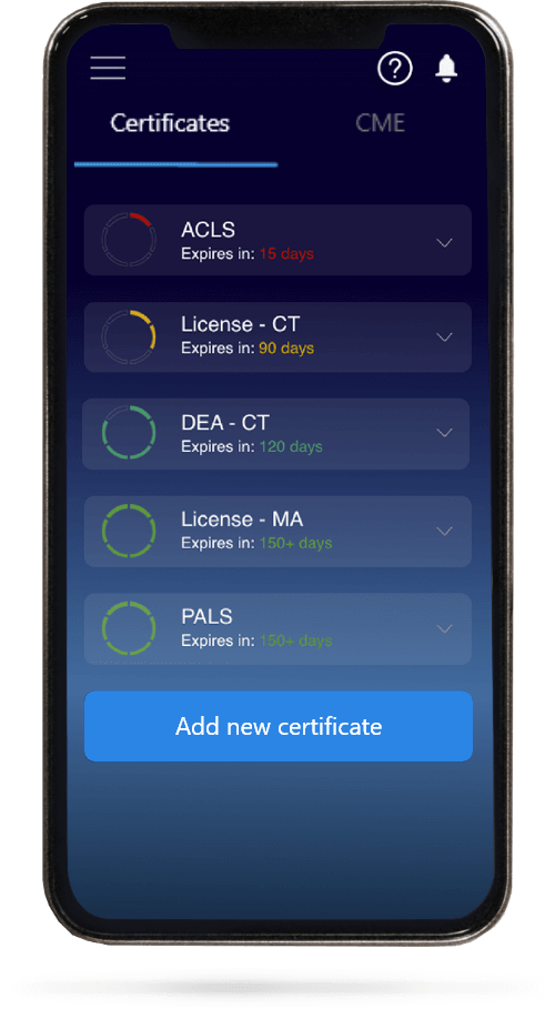
When you have your licenses, certificates and CMEs in one place, it's easier to track your career growth. You can easily share these with hospitals as well, using your medtigo app.